




Ⅰ. 서론
백악질골형성이상(COD, cemento osseous dysplasia)은 치근단 및 치아 지지부위의 골조직이 섬유조직과 화생골로 대체되는 골내병변으로[1] 중년여성에서 호발하고, 하악에서 많이 나타난다[2]. 원인은 명확히 알려지지 않았다[1].
2005년 WHO는 COD를 치근단백악질골형성이상(periapical COD), 국소형백악질골형성이상(focal COD), 개화성백악질골형성이상(florid COD)의 3가지 형태로 분류하였다[3,4]. 하악전치부에 여러 개 병소가 있는 경우는 치근단COD, 하악구치부에 한정적으로 나타나는 경우는 국소형COD, 상악과 하악구치부에서 다발적으로 나타나는 경우는 개화성COD로 명명하였다[5]. 여러 부위에서 나타나는 개화성COD는 치근단이나 국소형이 진행되어 확장된 병변으로 보기도 한다.
주로 하악구치부에서 나타나는 국소형COD는 특별한 증상 없이 우연히 발견되며[6] 일본, 한국 등 동아시아지역에서 많이 보고되었다 2) . 방사선사진에서는 비교적 명확한 경계를 가진 방사선투과상으로 관찰되기도 하고, 투과상내에 불투과상이 있는 혼합상으로 관찰되기도 한다[6].
방사선혼합상인 경우에는 골화섬유종(ossifying fibroma)으로 오진되어 외과적으로 제거하기도 하고, 특별한 증상이 없는 경우 그대로 악골 내에 남겨두기도 한다[2]. 방사선투과상으로 관찰된 경우에는 치근단병소로 오인되어 근관치료가 시행된 경우도 많다[7,8]. 국소형COD는 종양과는 달리 성장이 제한적이기에 특별한 치료가 요구되지 않는다. 그러므로 불필요한 치료가 진행되지 않도록 정확한 진단이 필요하다.
이에 저자는 치근단병소로 의심되었거나, 병소 제거가 요구되어 의뢰된 하악치아의 국소형 COD증례들을 보고하고, 임상의가 정확한 진단을 내릴 수 있도록 콘빔CT 영상을 공유하고자 한다.
Ⅱ. 증례
증례 1.
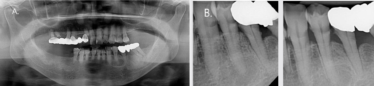
59세 여성 환자가 좌측 하악제1소구치 치근단낭 제거를 위해 본 원에 의뢰되었다. 임상검사에서 해당치아는 생활치로 나타났고, 타진과 저작시 이상반응이 없었다. 치근단과 파노라마방사선검사에서 병소의 경계는 치근단 방사선사진에서는 명확하지 않았고 파노라마사진에서는 명확하게 관찰되었다. 병소 내부는 주변골 보다는 투과상이나 완전한 방사선투과상으로 관찰되지 않았다. #34의 치주인대강과 치조백선은 정상이었고 치아변위나 치근흡수은 관찰되지 않았다(Fig. 1).
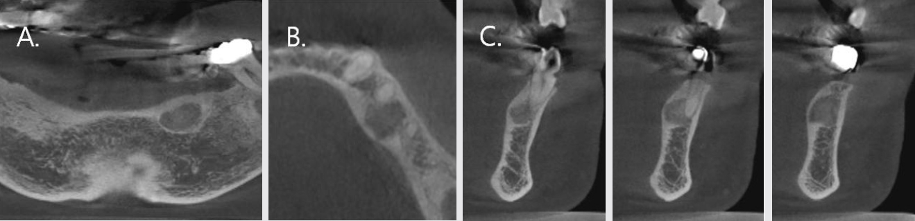
병소의 방사선투과성 정도와 치아와의 관계를 확인하기 위하여 콘빔CT검사를 시행하였다. 콘빔CT에서 경계가 명확하고 부분적으로 경화성 선으로 둘러싸인 방사선 투과상과 그 내부에 안개처럼 뿌연 불투과상이 존재하는 혼합상이 관찰되었다. 이 혼합상은 치근첨을 기준으로 대칭적이지 않았고, 원심과 설측으로 치우쳐져 있었다. 이로 인한 설측 피질골의 미약한 비박이 관찰되었으나 천공은 관찰되지 않았다(Fig. 2).
이와 같은 임상 및 방사선학적소견으로 증례 1은 치근단낭이 배제되었고, 하악소구치부에서 한정적으로 발생된 COD로 진단되었다. 향후 COD의 상태를 주기적으로 관찰하기로 하였다
증례 2.
48세 여성 환자가 신경치료 받았던 우측 하악대구치의 치근염증으로 발치 후 임플란트치료를 권유받았다. 그러나 발치 외에 다른 치료방법을 원해서 본 원에 내원하였다.
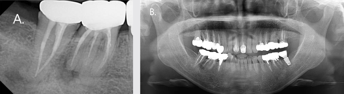
임상검사에서 해당 치아는 타진이나 저작시 이상소견이 없었다. 치근단 및 파노라마방사선검사에서 제1대구치, 제2대구치에서 비교적 명확한 경계를 가진 치근단주위 방사선투과상과 치근들과 중첩된 불투과상들이 함께 관찰되었다(Fig. 3).
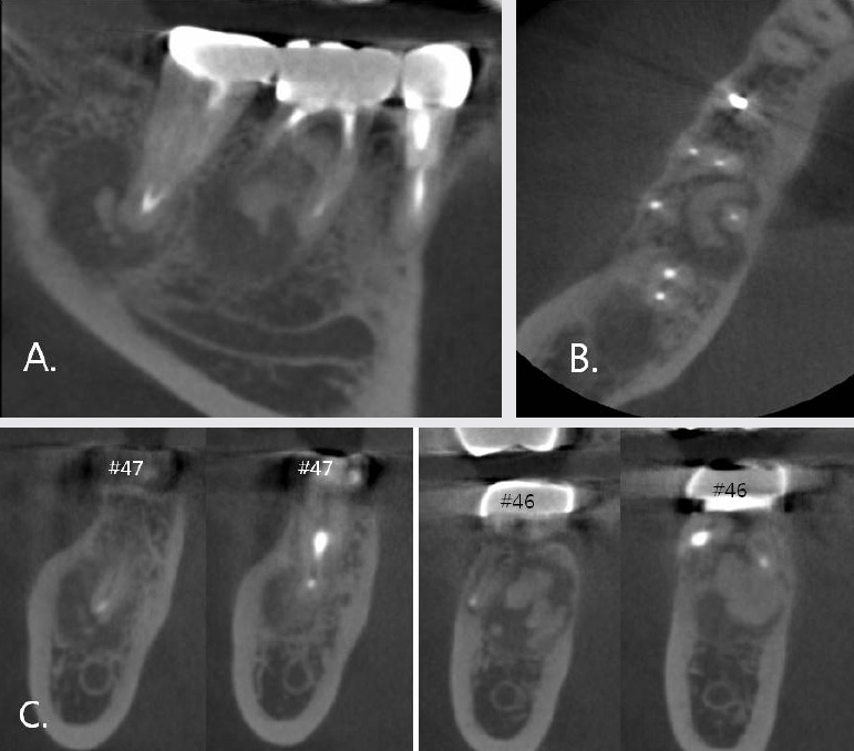
병소의 경계와 치근과 내부 불투과상의 관계를 알아보기 위하여 콘빔CT검사를 시행하였다. 대구치 치근단주위에서 경계가 명확한 방사선혼합상이 관찰되었다. 방사선 불투과상들은 치근과 분리되어 있었으며, 투과상내부에 있었다. 병소는 하악관 상부에 위치했고 치근첨을 기준으로 원심쪽에 치우쳐져 있었다, 제1대구치의 경우에는 불투과상이 설측에 주로 위치되어 있었고 그로 인한 설측 피질골 비박이 관찰되었다. 그러나 천공은 관찰되지 않았다(Fig. 4).
이상의 임상 및 방사선학적소견으로 증례 2는 하악구치부에서 한정적으로 발생된 COD로 진단되었다. 재신경 치료나 타 원에서 권유 받았던 발치를 시행하지 않고, COD의 상태를 주기적으로 지켜보기로 하였다.
증례 3.
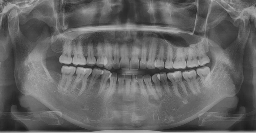
43세 여성 환자가 구강검진 중에 좌측 하악견치부위에서 방사선투과상이 발견되어 정확한 진단을 위해 본 원에 의뢰되었다. 문진이나 임상검사에서 특별한 증상을 호소하지 않았다. 파노라마방사선검사에서 좌측 하악견치와 제1소구치 치근단주위에 경계가 명확한 방사선투과상과 그 내부에 방사선불투과상들이 산재되어있는 혼합상이 관찰되었다(Fig. 5).
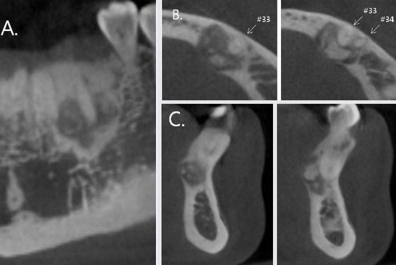
병소의 경계와 내부 상태, 해당 치아들과의 관계를 알아보고자 콘빔CT검사를 시행하였다. 견치와 제1소구치 치근주위에서 경화성 경계를 가진 방사선투과상과 내부에 다양한 크기의 불투과상들이 존재하는 혼합상이 관찰되었다. 불투과상들은 치근과 분리되었고 협측보다는 설측에서 주로 관찰되었다. 설측피질골의 팽윤과 비박이 관찰되었으나, 천공은 뚜렷하지 않았다. 견치의 치근이 미약하게 원심변위되어 제1소구치 치근과 가까워졌음이 관찰되었다(Fig. 6).
증례 3은 타 원에서 단순골낭 (simple bone cyst)을 의심하여 의뢰되었으나, 이상의 임상 및 방사선학적소견을 바탕으로 하악견치을 중심으로 한정적으로 발생된 COD로 진단되었다.
Ⅲ. 토의
악골의 섬유골성병변인 COD는 치근단백악질형성이상(periapical cemental dysplasia)으로 알려졌었으나, 2005년 WHO는 발생 위치를 기준으로 치근단, 국소형, 개화성의 3형태로 분류하였다[3,4]. COD는 섬유조직 내에 미성숙골, 백악질 등의 광물화 조직들이 혼합된 조직학적 양상을 가지며, 치주인대강에서 유래된 반응성병변이고 비종양성 성장의 특성을 가지고 있다[4].
중년 흑인 여성에 호발하고 해당 연령대에서 5.5%의 발병률을 보이지만[4] 전체 발병률은 0.3~0.4%로 비교적 흔한 병변은 아니다[9,10]. 보고된 형태별 발생 비율은 국소형이 16~52%, 개화성이 14.6%~65%로 다양한데[9~13], 그 이유는 병변이 존재해도 증상이 없는 경우가 많았고 방사선 불투과상이나 혼합상이 뚜렷하게 관찰되지 않은 경우들이 많았기 때문이라고 하였다[6]. 구치부에서 한정적으로 발생되는 COD는 한 동안 양성종양인 골화섬유종의 변이로 간주되었다가[5], 1994년 골형성이상 221 증례를 연구했던 Summerlin 등(1994) 6) 에 의해서 국소형COD라고 명명되었다. 대부분 증상 없이 우연히 발견되었고 남성보다는 여성에서, 하악제1대구치에서 가장 많이 발견되었다고 하였다[2,5,9]. 관련 치아는 치수감염과는 무관한 생활치이다[5]. 본 증례들도 이전 보고들처럼, 환자는 40~50대 중년 여성이 였고, 하악치아에 한정되어 있었으며, 특별한 임상증상이 없었다.
COD 진단은 조직검사를 시행하지 않고 임상 및 방사선학적소견만으로 이루어진다[4]. 방사선학적소견이 애매한 경우에, 치수생활력검사는 국소형COD와 치수감염에 의한 치근단병소를 감별하는데 도움을 준다. 많은 보고에서 방사선검사의 중요성을 강조하였다[7,8,10,14]. 대부분 병소는 명확한 경계를 가진 방사선불투과상이나 혼합상으로 관찰되었다. 병소에 의한 치아 변위는 드물며[10], 치조 백선과 치주인대강은 정상이다[13]. 콘빔CT는 일반방사선 사진에서 관찰되지 않는 병소의 협·설 관계, 병소의 방사선불투과성 정도를 명확하게 보여줌으로써 COD를 진단하는데 많은 도움을 준다[2,15~17]. 콘빔CT는 일반CT보다 적은 방사선량으로 여러 방향에서 병소를 관찰할 수 있게 해주고, 중첩된 상이 아닌 원하는 단면에서 병소의 위치나 크기를 보다 명확하게 보여주기 때문이다[16]. 그리고 병소 내부의 석회화 정도 2) , 주변 구조물인 협·설 피질골의 팽윤이나 천공 여부를 명확하게 보여준다[15~17]. COD에서는 피질골의 변화 중 비박이 제일 많이 관찰된다[10,17]. 본 증례들도 치근단과 파노라마 영상에서 명확하지 않았던 병소의 내부 석회화 정도, 치근과 불투과상과의 관계, 치아에 대한 병소의 협·설 관계, 피질골의 비박 및 팽윤을 콘빔CT 영상에서 확인한 후 치근단염증이나 낭을 배제할 수 있었다.
초기에는 섬유 조직이 였다가 석회화가 점점 증가하는 성숙단계를 거치게 되는 COD의 특성 때문에 특징적인 방사선학적소견 없이, 방사선투과상, 혼합상, 불투과상이 다양하게 관찰된다. 성숙단계에서 병소내부 석회화에 의한 방사선불투과상이 나타나야 함에도 불구하고, 실제 방사선불투과상으로 관찰된 경우도 의외로 적었다[6]. 이런 경우, 임상가들은 종종 COD보다는 염증이나 낭등의 치근단병소로 오인하게 된다[5]. COD를 치근단염이나 낭으로 오진하여, 불필요한 근관치료가 시행되었던 경우가 각각 21.7% [10], 7.3% [17] 였다는 보고들이 있으며, 모두 정확한 진단의 중요성을 강조하였다[7,8,10,14].
그러므로, 중년 여성의 구치부에서 뚜렷한 방사선투과상이 아닌 치근단주위 방사선혼합상이 의심될 때, 치수의 실활 여부 확인과 더불어 콘빔CT 영상에서 병소내부의 방사선불투과상 정도, 피질골 변화를 확인하는 것이 중요하다. 또한, 콘빔CT는 COD의 진단 뿐 만 아니라 COD의 주기적 관찰이 필요할 때 가장 추천되는 방사선검사이다[15].


 PDF Links
 PubReader
 ePub Link
 XML Download
 Full text via DOI
 Download Citation
 Print
Print


