구강 스캐닝과 전통적 인상 채득의 작업 시간 및 환자 불편감 비교: 체계적 문헌 고찰 및 메타분석

Intraoral scanning versus conventional impressions for working time and patient discomfort: A systematic review and meta-analysis

Article information

J Korean Dent Assoc. 2026;64(7):223-234
Publication date (electronic) : 2026 July 31
doi : https://doi.org/10.22974/jkda.2026.64.7.001
1Department of Computer Graphics Technology, Polytechnic Institute, Purdue University, West Lafayette, USA
2Edeun Union Dental Clinic, Busan, Korea
*Corresponding author: Dr. Jaehoon Cho Edeun Union Dental Clinic, 403, 4F, M-Tower, 469-1, Yeonsan-dong, Yeonjegu, Busan 47569, Korea Tel: +82-51-791-0002, E-mail: chojae716@naver.com
Received 2026 March 3; Revised 2026 June 8; Accepted 2026 June 15.

Trans Abstract

Purpose

Intraoral scanning (IOS) is increasingly used as a digital alternative to conventional impressions, yet reported benefits for chairside efficiency and patient experience vary across studies. This review quantified differences in working time and patient-reported discomfort and explored effects by age and conventional impression material.

Materials and Methods

PubMed, the Cochrane Library, Google Scholar, and RISS were searched in accordance with PRISMA 2020 for randomized or prospective comparative studies. Random-effects meta-analyses pooled mean differences (MD) for working time and Hedges’ g standardized mean differences (SMD) for discomfort on visual analog scales. Prespecified subgroup analyses were conducted by age group and conventional impression material.

Results

Fifteen studies were included. IOS shortened working time (MD −3.39 min; 95% CI −5.16 to −1.62), with larger time savings versus polyvinyl siloxane and polyether than alginate. IOS also reduced discomfort (SMD −1.24; 95% CI −1.53 to −0.94), with consistent benefits across materials and across adult and pediatric populations.

Conclusion

IOS improves patient comfort and modestly reduces chairside time compared with conventional impressions. These findings support IOS use in routine clinical practice and suggest that time reductions are more pronounced when conventional impressions use polyvinyl siloxane or polyether rather than alginate

서론

최근 치과 진료 환경은 CAD/CAM 기술의 발전과 함께 디지털 워크플로를 중심으로 빠르게 변화하고 있다. 이러한 변화의 핵심 요소 중 하나인 구강 스캐닝(intraoral scanning, IOS)은 전통적인 인상 채득 과정을 디지털 데이터로 대체함으로써, 보철 및 수복 치료 전반에서 활용 범위를 지속적으로 확대해왔다. 구강 스캐너는 구강 내 구조를 직접 촬영하여 3차원 디지털 인상을 획득할 수 있게 하며, 이는 인상재와 트레이를 사용하는 기존 방식에서 발생할 수 있는 물리적·기계적 한계를 보완하는 기술로 평가받고 있다.

기존의 전통적인 인상 채득 방식은 alginate나 실리콘 기반 인상재를 사용하여 인상을 채득하고 경화가 완료될 때까지 구강 내에 유지해야 하는 특성상, 환자에게 불편감, 구역 반사, 불쾌한 맛, 호흡 곤란 등의 문제를 유발할 수 있다[1]. 이러한 과정은 환자의 협조도에 영향을 미칠 뿐 아니라, 술자 입장에서도 인상 실패로 인한 재채득, 진료 시간 증가, 작업 효율 저하로 이어질 가능성이 있다[2]. 반면 구강 스캐닝은 인상재 사용을 최소화하거나 제거함으로써, 환자의 신체적 부담을 줄이고 보다 간소화된 임상 프로토콜을 제공할 수 있는 대안으로 제시되고 있다[3].

구강 스캐닝과 전통적 인상 채득 방식의 비교에 관한 기존 연구들은 정확도, 정밀도, 변연 적합도 등 기술적 측면을 중심으로 다수 보고되어 왔으며, 두 방법 간 임상적으로 허용 가능한 수준의 결과를 보인다는 점이 반복적으로 제시되어 왔다[4]. 최근에는 환자 선호도, 만족도, 불편감과 같은 환자 보고 결과(patient-reported outcome measures, PROMs)에 대한 관심도 점차 증가하고 있다[5]. 그러나 이러한 연구들은 특정 임상 상황이나 제한된 결과 변수에 초점을 맞춘 경우가 많아, 결과 해석에 있어 일관성이 부족하다는 한계를 지닌다. 또한 연구 설계, 사용된 스캐너와 인상재의 종류, 평가 방식의 차이로 인해 이질성이 크며, 인상 채득 시간과 환자 불편감이 부차적 결과로 다뤄지거나 체계적으로 비교되지 않은 경우도 적지 않다.

이에 본 연구는 기존 문헌에서 제기된 이러한 한계를 보완하고자, 구강 스캐닝과 전통적 인상 채득 방식 간의 차이를 인상 채득 시간과 환자 불편감이라는 임상적으로 중요한 두 가지 변수에 초점을 맞추어 분석하고자 한다. 특히, 체계적 문헌 고찰 및 메타분석을 통해 두 인상 방식의 임상적 특성을 종합적으로 비교함으로써, 임상 환경에서 보다 효율적이고 환자 중심적인 인상 채득 방법 선택에 근거를 제공하는 것을 본 연구의 목적으로 한다.

재료 및 방법

본 연구는 체계적 문헌 고찰 및 메타분석(systematic review and meta-analysis)으로, Preferred Reporting Items for Systematic Reviews and Meta-Analyses(PRISMA) 2020 가이드라인에 의거하여 수행되었다. 연구의 프로토콜은 사전에 정의된 Population, Intervention, Comparison, Outcome, Study Design(PICOS) 전략에 따라 수립되었다.

문헌 검색은 2025년 11월 1일부터 2025년 11월 30일까지 PubMed, Google Scholar, Cochrane Library, RISS 등의 국내외 데이터베이스를 이용하여 진행되었다. 문헌 검색에는 구강 스캐너, 디지털 인상, 전통적 인상, 작업 시간, 환자 불편감, visual analogue scale(VAS) 등 MeSH 기반 용어를 조합하여 사용하였다. 실제 수행한 상세 검색 전략은 다음과 같다. ("intraoral scanner" or “digital impression”) and ("conventional impression") and ("working time" or "patient discomfort”).

문헌의 선정 및 배제 과정은 PICOS criteria에 기반하여 수행되었다. 이에 따른 구체적인 포함 기준과 배제 기준은 Table 1에 상세히 기술하였다.

Inclusion and exclusion criteria (PICOS framework)

본 연구는 사람을 대상으로 구강 스캐닝과 전통적 인상채득을 직접 비교한 무작위 대조 임상시험 및 전향적 대조 비교 임상연구를 포함하였다. 대상자 포함 기준은 진단 또는 고정성 보철물 제작을 위해 인상 채득이 필요한 환자로 설정하였다. 증례보고, in vitro 연구, 종설, 학위논문 등은 제외하였다.

문헌 선택 과정은 두 명의 연구자가 독립적으로 수행하였으며, 1차적으로 제목과 초록을 스크리닝하여 관련 없는 문헌을 배제하였고 2차적으로 원문을 검토하여 최종 포함 여부를 결정하였다. 선정 과정에서 연구자 간의 의견 불일치가 발생한 경우에는, 해당 문헌을 재검토하고 상호 간의 충분한 논의를 거쳐 최종 합의에 도달하였다. 문헌 선택의 전체 과정은 PRISMA flow diagram를 통해 제시하였다(Fig. 1).
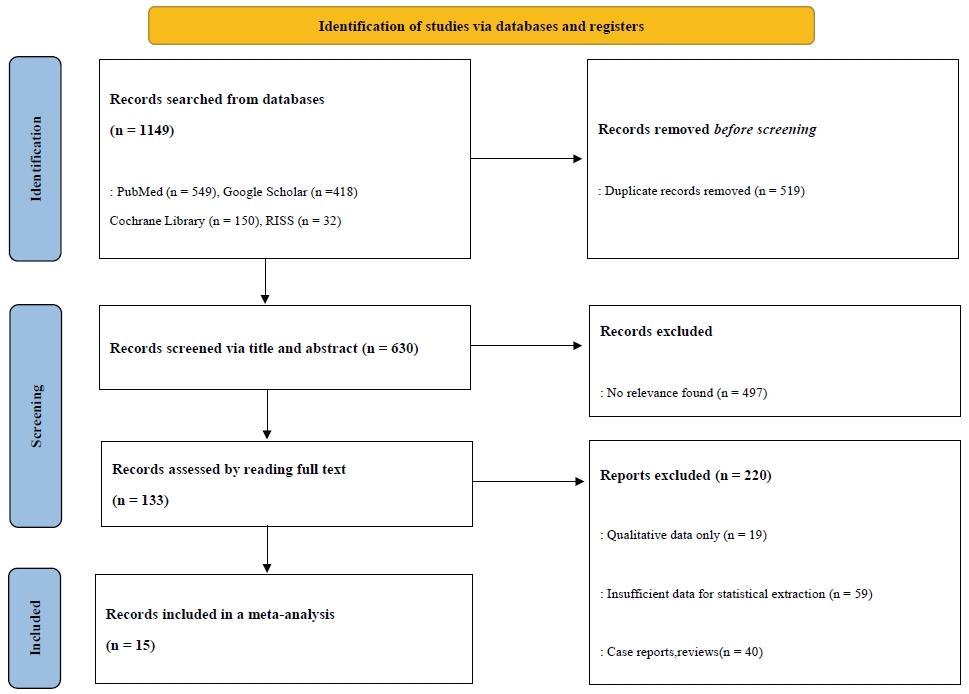
Fig. 1.

PRISMA 2020 flow diagram depicts the study identification, screening, eligibility assessment, and inclusion process for the systematic review and meta-analysis comparing intraoral scanning with conventional impression techniques.

최종 선정된 문헌에서는 제1저자, 출판 연도, 대상자 수, 환자의 평균 연령(성인/소아), 사용된 구강 스캐너의 종류, 대조군 인상재의 종류(alginate, polyvinyl siloxane, polyether), 그리고 각 그룹에서 인상채득에 소요된 작업시간 및 VAS의 평균과 표준편차 등을 추출하였다. 본 연구는 인상 채득 방식 자체의 효율성을 비교하기 위해 체어사이드에서 이뤄진 실제 작업 시간만을 분석 범위로 하였다. 따라서 기공 과정을 포함한 denture delivery 등 전체 워크플로우에 사용된 시간은 정량적 합성에서 제외하였다.

포함된 연구의 비뚤림 위험 평가는 Cochrane Risk of Bias(RoB) 2.0의 5가지 도메인 구조를 기반으로 수행하였다. 각 도메인은 D1(무작위 배정 과정에서 발생하는 비뚤림), D2(의도한 중재에서 이탈함으로 인한 비뚤림), D3(결측 결과 데이터로 인한 비뚤림), D4(결과 측정의 비뚤림), D5(보고된 결과 선택의 비뚤림)을 의미한다.

다만 일부 전향적 비교연구(비무작위 연구)의 경우 무작위배정 과정에 관한 도메인 해석에 제한이 있어, 해당 연구 설계에서 기인할 수 있는 교란 및 선택편향 가능성을 결과 해석에서 보수적으로 반영하였다.

메타분석은 R software (ver. 4.5.2, R Foundation for Statistical Computing, Vienna, Austria)의 'meta' 패키지를 사용하여 수행되었다. 작업 시간은 측정 단위가 동일하므로 평균 차(mean difference, MD)를, 환자 불편감은 연구마다 척도의 범위가 다를 수 있음을 고려하여 표준화된 평균 차(standardized mean difference, SMD)를 효과크기로 산출하였으며, 95% 신뢰구간을 함께 제시하였다. 연구 간의 이질성은 I2 통계량과 Cochrane's Q test를 통해 평가하였다. 본 연구에 포함된 문헌들은 구강 스캐너의 종류, 술자의 숙련도, 그리고 대조군 인상재의 물성 등 임상적 이질성이 존재한다고 판단되었기에, 보수적인 추정을 위하여 무작위 효과 모형(random-effects model)을 적용하였다. 또한, 이질성의 원인을 규명하기 위해 환자의 연령과 인상재의 종류에 따른 하위그룹 분석을 시행하였다. 출판 편향은 funnel plot의 시각적 대칭성을 확인하고 Egger's 회귀검정을 통해 통계적 유의성을 검증하였다. 모든 통계적 유의수준은 p < 0.05로 설정하였다.

결과

문헌 선정의 전 과정은 PRISMA flow diagram 으로 도식화하였다(Fig. 1). 데이터베이스 검색 및 중복 문헌 제거 후, 제목/초록 선별과 원문 평가를 거쳐 최종적으로 15편의 연구가 메타분석에 포함되었다(Table 2) [6-20]. 최종 선정된 15편의 문헌 중,체어사이드 작업 시간에 부합하는 정량자료(분 단위, mean ±SD)를 보고하여 메타분석에 포함 가능한 무작위 대조 임상시험(randomized controlled trial, RCT)은 13편이었으며, 나머지 2편은 전향적 대조 비교 연구였다. 한편, VAS를 기반으로 환자 불편감을 정량자료로 보고하여 메타분석에 포함 가능한 문헌은 14편이었다.
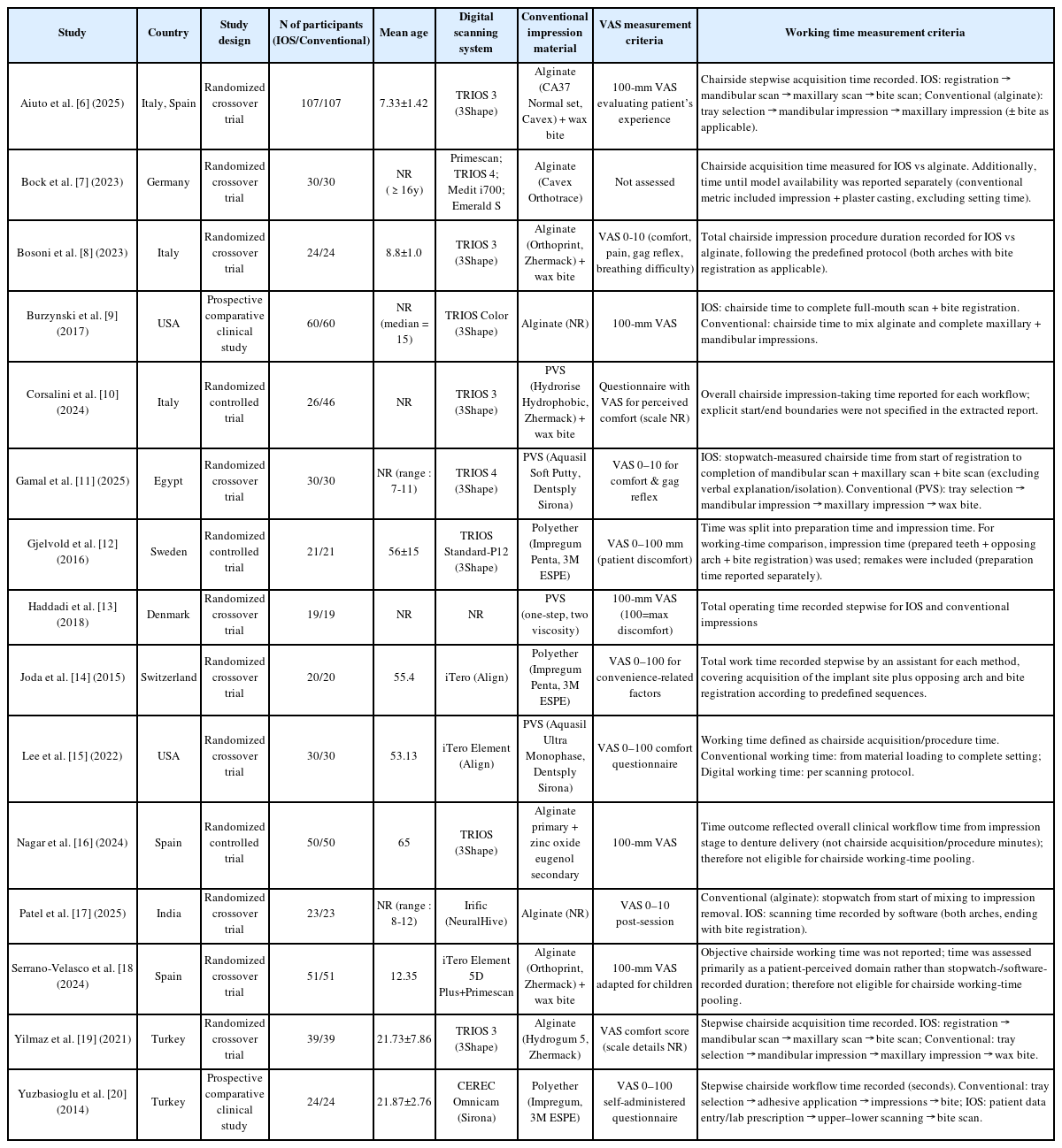
General characteristics of included studies

ROB 2.0에 따라 15편의 포함 연구를 평가한 결과, 전반적 비뚤림 위험은 Some concerns 11편(73.3%), High risk 4편(26.7%)으로 분류되었다. 도메인별 분포는 다음과 같다. 무작위화 과정(D1)은 Low 3편(20.0%), Some concerns 9편(60.0%), High 3편(20.0%)이었다. 의도한 중재에서의 이탈(D2)은 Low 10편(66.7%), Some concerns 5편(33.3%)으로 분류되었다. 결측 결과자료(D3)는 Low 10편(66.7%), Some concerns 4편(26.7%), High 1편(6.7%)이었다. 결과 측정(D4) 및 보고된 결과 선택(D5)은 모든 연구에서 Some concerns(각 15편, 100%)으로 평가되었다(Figs. 2 and 3).
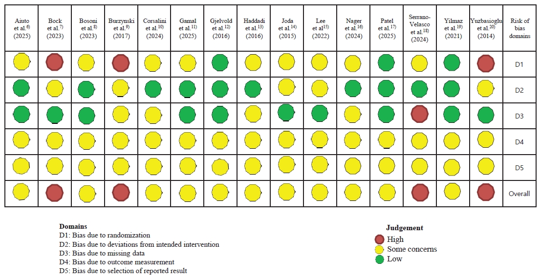
Fig. 2.

Risk of bias (RoB 2.0) traffic-light plot for the included studies. Domain-level judgements are presented for D1 (randomization process), D2 (deviations from intended interventions), D3 (missing outcome data), D4 (measurement of the outcome), and D5 (selection of the reported result), along with the overall RoB 2.0 judgement for each study.
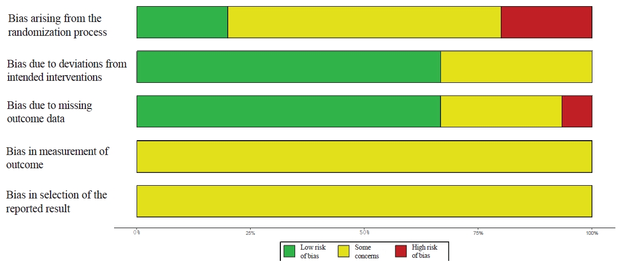
Fig. 3.

Summary plot of Risk of bias (RoB 2.0) judgements shows the proportion of included studies rated as low RoB 2.0, some concerns, or high risk of bias for each RoB 2.0 domain (D1–D5) and the overall judgement.

메타분석 결과: 작업 시간

작업시간은 13편 연구(구강 스캐닝 453명, 전통적 인상채득 473명)를 대상으로 랜덤효과모형으로 분석하였다. 전체 효과는 MD = −3.39 minutes(95% CI, −5.16 to −1.62 ; p < 0.001)로, 디지털 구강 스캐닝이 전통적 인상채득에 비해 유의하게 작업시간을 단축시키는 것으로 나타났다. 다만 이질성은 높았으며(I² = 99.5%, τ² = 10.3519, Q 검정 p < 0.001), 이에 따라 환자 연령군과 인상재에 따른 하위그룹 분석을 진행하였다.

환자 연령군에 따른 하위그룹 분석 결과는 Figure 4와 같다. 성인군은 MD = −3.54 minutes(95% CI, −6.14 to −0.94), 소아군은 MD = −3.10 minutes(95% CI, −3.97 to −2.23) 로 두 그룹에서 모두 구강 스캐닝이 전통적 인상채득에 비해 작업시간을 유의하게 단축시키는 것으로 확인되었다. 점추정치 기준으로 성인군에서 소아군 대비 더 큰 효과크기가 관찰되었으나, 연령군에 따른 하위그룹별 효과 크기의 차이는 유의하지 않았다(χ² = 0.10, df = 1, p = 0.7523).
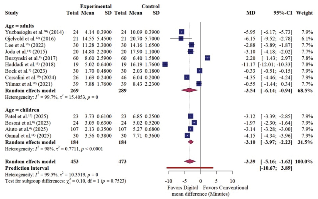
Fig. 4.

Forest plot of working time (minutes) comparing intraoral scanning (IOS) versus conventional impression techniques, stratified by age group (adults vs children), using a random-effects model. Effect sizes are presented as mean differences (MDs) with 95% confidence intervals (CIs); negative MDs values favor IOS (shorter working time). Square sizes reflect study weights and diamonds indicate pooled estimates (with a prediction interval where shown).

인상재 유형에 따른 하위그룹 분석 결과는 Figure 5와 같다. 하위그룹별 효과 크기에 있어 통계적으로 유의한 차이가 확인되었다(χ² = 10.06, df = 2, p = 0.0065). 구체적으로, 구강 스캐닝은 polyvinyl siloxane과 비교하였을 때 MD = −5.63 minutes(95% CI, −9.29 to −1.98)로 작업시간에서 가장 큰 단축 효과를 나타냈다. 한편, polyether와 대비해서는 MD = −4.91 minutes(95% CI, −6.99 to −2.82)로 작업시간 단축 효과가 뚜렷하였다.
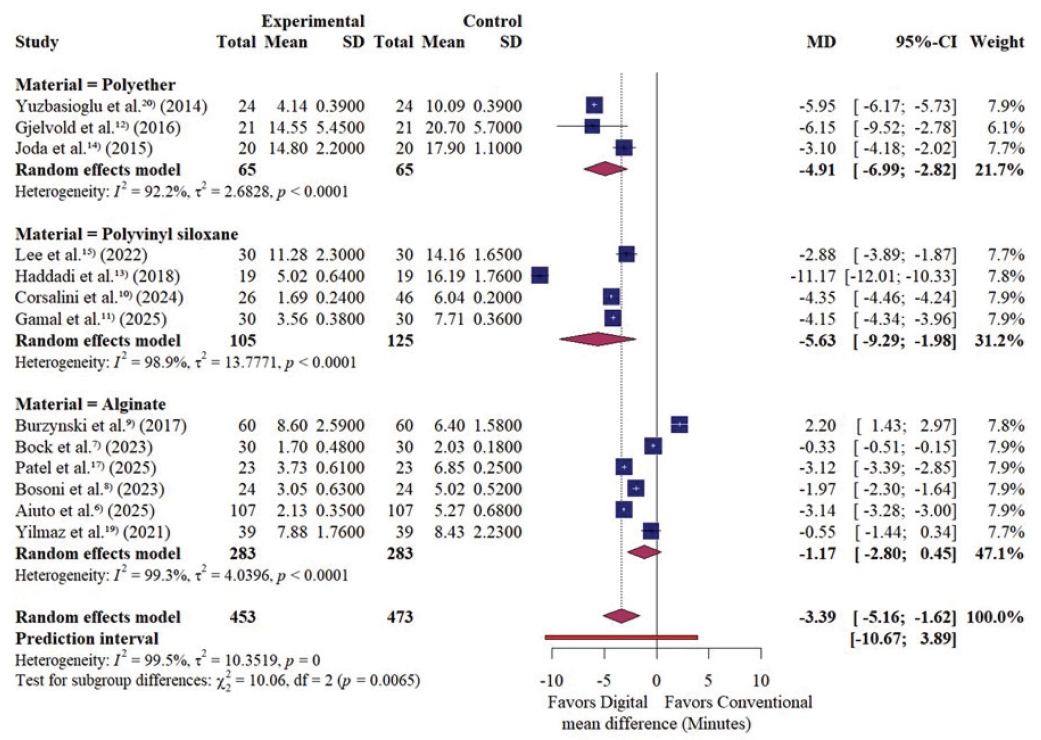
Fig. 5.

Forest plot of working time (minutes) by conventional impression material (polyether, polyvinyl siloxane, and alginate) comparing intraoral scanning (IOS) versus conventional impressions using a random-effects model. Effect sizes are presented as mean differences (MDs) with 95% confidence intervals (CIs); negative mean difference values favor IOS. Square sizes reflect study weights and diamonds represent pooled estimates (with a prediction interval where shown).

반면 alginate 하위그룹은 MD = −1.17 minutes(95% CI, −2.80 to 0.45)로 나타났다. 이는 구강 스캐닝이 작업시간을 단축시키는 경향성을 보였으나, 통계적으로 유의미한 차이는 관찰되지 않았음을 의미한다.

메타분석 결과: 환자 불편감

환자 불편감은 14편 연구(구강 스캐닝 524명, 전통적 인상 채득 544명)에서 추출되었으며, 연구 간 척도/분산 차이를 고려하여 SMD 기반으로 랜덤효과모형 분석을 수행하였다. 전체 효과는 SMD = −1.24(95% CI, −1.53 to −0.94) 로, 구강 스캐닝이 전통적 인상채득에 비해 환자 불편감을 유의하게 감소시키는 것으로 나타났다. 이질성은 중등도–높은 수준이었다(I² = 80.8%, τ² = 0.2398, Q 검정 p < 0.001). 이에 따라 환자 연령군과 인상재에 따른 하위그룹 분석을 진행하였다.

연령군 하위그룹 분석에서 성인 그룹은 SMD = −1.29(95% CI, −1.70 to −0.87), 소아 그룹은 SMD = −1.16(95% CI, −1.58 to −0.74)으로 두 군 모두 구강 스캐닝이 전통적 인상채득 대비 불편감을 유의하게 감소시키는 것으로 나타났다(Fig. 6). 점추정치 기준으로 성인 그룹에서 더 큰 효과크기가 수치상으로 관찰되었으나, 연령군에 따른 하위그룹별 효과 크기의 차이는 통계적으로 유의하지 않았다(χ² = 0.17, df = 1, p = 0.6767).
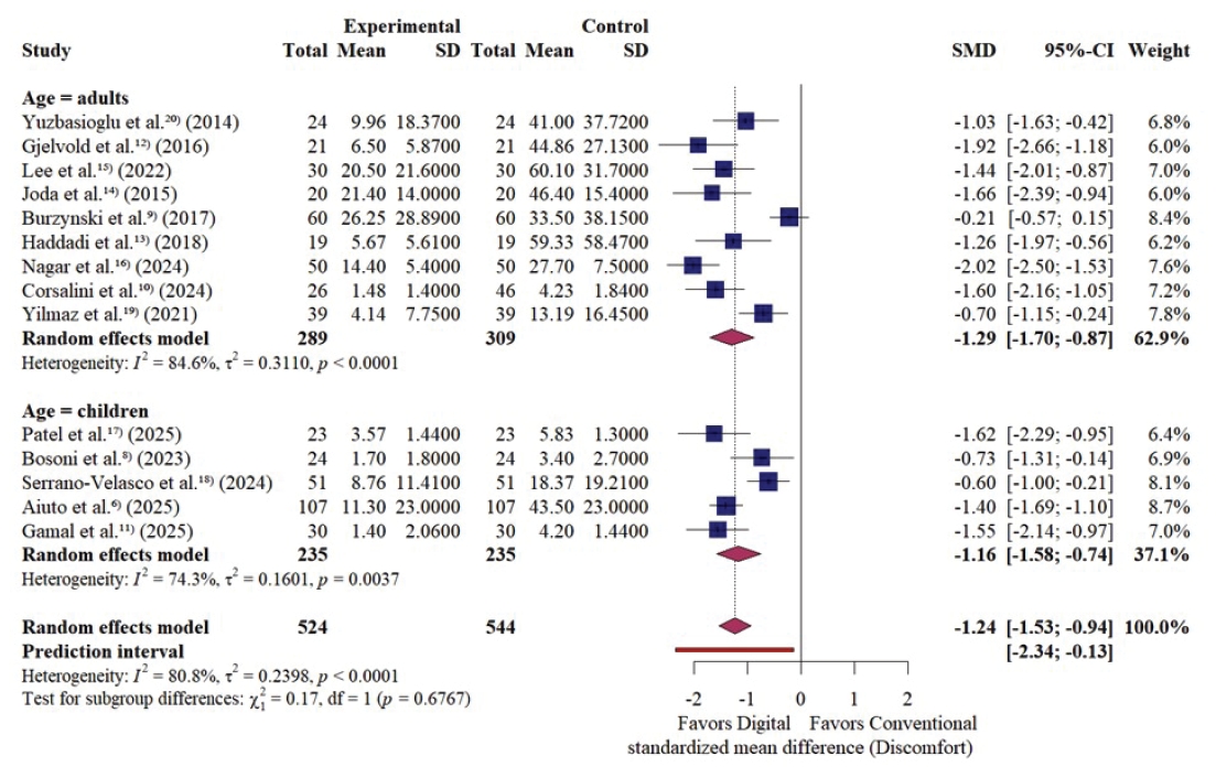
Fig. 6.

Forest plot of patient discomfort comparing intraoral scanning (IOS) versus conventional impression techniques, stratified by age group (adults vs children), using an inverse variance (IV) random-effects model. Effect sizes are presented as standardized mean differences (SMDs) with 95% confidence intervals (CIs); negative SMDs values favor IOS (lower discomfort). Square sizes reflect study weights and diamonds indicate pooled estimates (with a prediction interval where shown).

인상재 기준 하위그룹 분석에서, polyether는 SMD = −1.50(95% CI, −2.04 to −0.96), polyvinyl siloxane은 SMD = −1.49(95% CI, −1.78 to −1.19), alginate는 SMD = −1.03(95% CI, −1.51 to −0.54)로 모든 하위그룹에서 구강 스캐닝이 전통적 인상채득 대비 불편감을 유의하게 감소시키는 것으로 나타났다(Fig. 7). 점추정치 기준으로는 polyether, polyvinyl siloxane, alginate 순으로 높은 효과크기가 수치상으로 관찰되었으나, 인상재에 따른 하위그룹별 효과크기의 차이는 유의하지 않았다(χ² = 2.77, df = 2, p = 0.2503).
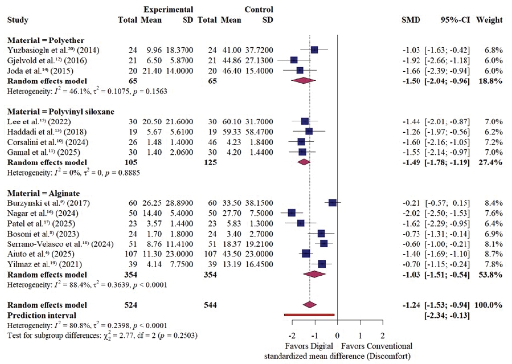
Fig. 7.

Forest plot of patient discomfort by conventional impression material (polyether, polyvinyl siloxane, and alginate) comparing intraoral scanning (IOS) versus conventional impressions using an inverse variance (IV) random-effects model. Effect sizes are presented as standardized mean differences (SMDs) with 95% confidence intervals (CIs); negative SMD values favor IOS. Square sizes reflect study weights and diamonds represent pooled estimates (with a prediction interval where shown).

출판편향(publication bias) 평가

funnel plot 시각적 평가에서 작업시간 및 불편감 결과 모두에서 뚜렷한 비대칭은 관찰되지 않았다(Figs. 8 and 9). Egger’s 회귀검정 결과, 작업시간은 t = 0.37, df = 11, p = 0.7156, 환자 불편감은 t = −1.49, df = 12, p = 0.1630으로 두 결과 모두에서 funnel plot의 비대칭을 시사하는 통계적 근거는 확인되지 않았다. 다만, 포함된 연구 수가 제한적이므로 출판편향 가능성을 완전히 배제할 수는 없으며, 해석에 주의가 필요하다.
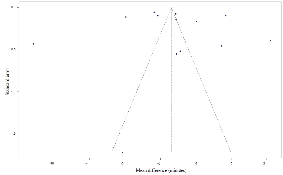
Fig. 8.

Funnel plot assessing potential small-study effects/publication bias for the working time outcome comparing intraoral scanning with conventional impression techniques. Each point represents an individual study plotted by effect size (mean difference, minutes) against its standard error; the vertical line indicates the pooled effect estimate and the dashed lines indicate pseudo 95% confidence limits.
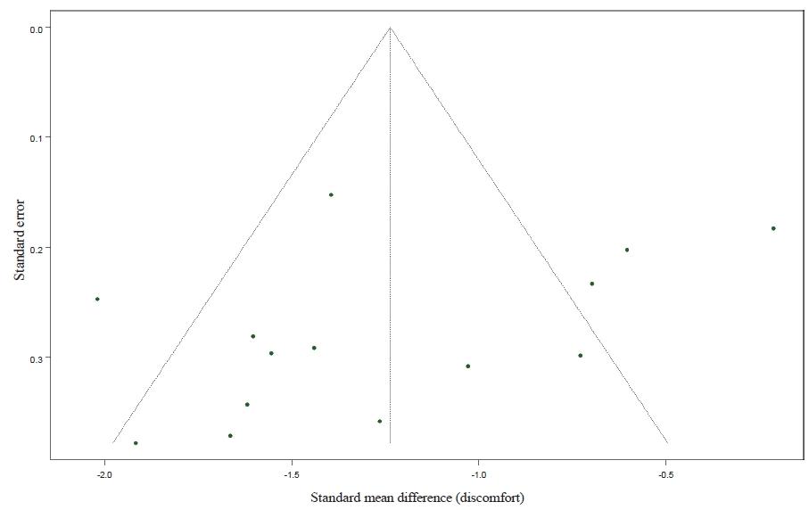
Fig. 9.

Funnel plot assessing potential small-study effects/publication bias for the patient discomfort outcome comparing intraoral scanning with conventional impression techniques. Each point represents an individual study plotted by effect size (standardized mean difference) against its standard error; the vertical line indicates the pooled effect estimate and the dashed lines indicate pseudo 95% confidence limits

고찰

본 체계적 문헌고찰 및 메타분석은 구강 스캐닝과 전통적 인상 채득 간의 임상적 차이를 작업시간과 환자 불편감이라는 환자 중심 지표에 초점을 맞추어 종합적으로 평가하였다. 본 연구에서 구강 스캐닝은 전통적 인상 채득에 비해 작업시간을 평균 3.39분 단축시켰으며(MD = −3.39 min), 환자 불편감을 유의하게 감소시키는 것으로 나타났다(SMD = −1.24). 이러한 결과는 디지털 인상 채득이 환자 경험을 개선하면서 임상 효율성을 향상시킬 수 있다는 기존 임상 연구 및 체계적 문헌고찰 연구의 결론과 일치한다[14,21-23].

환자 불편감의 감소는 구강 스캐닝이 인상 트레이와 인상재를 구강 내에 장시간 유지해야 하는 과정을 제거함으로써, 구역 반사, 호흡 불편, 불쾌감(맛·냄새) 및 압박감을 줄이는 기전으로 설명될 수 있다[12,20]. 실제로 다수의 RCT에서 환자들은 디지털 인상 채득을 더 선호하거나 불편감이 더 낮다고 보고하였으며[12-14,20,21], 이는 본 연구의 전체 효과크기와 일치하는 결과라고 할 수 있다. 한편, Cohen은 표준화된 평균차에 대해 0.2/0.5/0.8을 각각 작은/중간/큰 효과로 해석하는 경험적 기준을 제시한 바 있다[24]. 따라서 본 연구에서 관찰된 환자 불편감의 SMD가 0.8을 상회한다는 점은, 구강 스캐닝의 전통적 인상채득 대비 불편감을 감소시키는 효과가 통계적으로 유의할 뿐 아니라 임상적으로도 유의미하다는 가능성을 시사한다.

작업시간의 단축은 구강 스캐닝이 인상재 혼합, 트레이 장착, 경화 대기 및 인상체 제거 과정에 소요되는 시간을 줄일 수 있다는 점에서 타당하다[12,13]. 특히 전통적 인상 채득에서 재인상 발생이나 술자·환자 협조도 문제로 인한 추가 시간이 발생할 수 있다는 점을 고려하면, 단순 평균 시간 차 이상으로 체감 효율성이 확대될 여지도 있다[20,22]. 다만 작업시간은 연구마다 정의가 상이하여(트레이 선택 및 접착제 도포 시간 포함 여부, 스캔 재촬영 포함 여부, 교합 채득 포함 여부 등), 본 연구의 추정치는 평균적인 경향으로 해석하는 것이 적절하리라 사료된다.

본 연구에서 작업시간 결과의 이질성이 매우 높게 관찰되었는데, 이는 작업시간의 정의와 측정 범위가 연구마다 완전히 동일하지 않았기 때문일 가능성이 크다. 이외에도 치과 디지털 장비 연구의 특성상 스캐너 세대/모델, 스캔 전략, 술자 숙련도, 보철·교정 등 임상 상황, 인상 범위(부분/전악) 가 복합적으로 작용하기 때문으로 해석된다[23,25,26]. 특히 구강 스캐닝은 술자 경험에 따라 스캔 품질과 소요 시간이 달라질 수 있고, 초기 러닝커브 구간에서는 오히려 시간이 증가할 수 있다는 점이 보고되어 왔다[26]. 반면, 전통적 인상채득은 재료 특성 및 술자 습관에 따라 비교적 프로토콜 시간이 고정적으로 형성되는 경향이 있어, 연구 간 작업시간 분산이 구강 스캐닝에서 더 크게 나타날 수 있다[25,26].

인상재 종류에 따른 하위그룹 분석에서 구강 스캐닝의 작업시간 단축 효과는 polyvinyl siloxane 및 polyether 대비 더 크게 나타났으나, alginate와 비교시에는 통계적으로 유의한 시간 단축 효과가 나타나지 않았으므로 임상 적용시 이를 고려해야 한다. 이는 polyvinyl siloxane/polyether의 경우 인상 채득 전 준비 단계가 많고(트레이 준비, 혼합 및 주입, 경화 대기 등) 구강 내 유지시간이 길어질 수 있는 반면, alginate는 상대적으로 간단하고 경화 시간이 짧아 시간 격차가 축소되기 때문으로 설명 가능하다[9,12,13]. 실제로 alginate와 구강 스캐닝을 직접 비교한 무작위 교차 연구에서도 시간 단축 효과는 상황, 측정 방식에 따라 결과가 상이할 수 있음이 보고된 바 있다[9,19]. 반면 불편감은 인상재 종류에 관계없이 구강 스캐닝이 일관되게 유리하였는데, 이는 인상재 종류와 무관하게 트레이 및 인상재 자체가 유발하는 구역 반사, 압박감 등의 불편 요인이 공통적으로 존재하기 때문으로 해석된다[22].

연령(성인/소아) 하위그룹 분석에서는 두 결과 모두에서 구강 스캐닝의 효과가 일관되었고, 하위그룹 간 차이는 유의하지 않았다. 이는 소아에서도 구강 스캐닝이 전통적 인상 대비 불편감을 줄이고 환자 선호를 향상시킬 수 있다는 기존 연구 결과와 일치한다[8,19]. 특히 소아·청소년에서는 구역 반사 및 불안이 인상 채득의 주요 장애 요인이 될 수 있는데, 본 연구 결과는 구강 스캐닝이 이러한 장벽을 낮출 수 있다는 임상적으로 중요한 가능성을 시사한다.

기존 연구는 디지털 인상 채득의 정확도, 적합도, 재현성 등 기술적 측면에 집중한 경우가 많았으나, 최근에는 PROMs와 작업 효율성의 중요성이 강조되고 있다[23]. Gallardo 등(2017) [22]은 체계적 문헌고찰을 통해 디지털 인상이 환자 경험 측면에서 우수하며, 작업시간은 연구 조건에 따라 유사하거나 디지털이 유리할 수 있다고 보고한 바 있다. 본 연구는 여기서 한 걸음 나아가, 환자 불편감과 작업시간을 핵심 지표로 설정하여 메타분석을 진행하였으며, 연령 및 인상재 종류에 따른 하위그룹 분석을 통해 임상적 해석 가능성을 확장했다는 점에서 의의를 갖는다. 또한 메타분석에 포함된 연구의 비뚤림 위험을 RoB 2.0으로 체계적으로 평가하고, 출판편향을 함께 검토함으로써 결과의 신뢰도를 다각도로 점검하였다.

본 연구의 한계는 다음과 같다. 첫째, 대상 문헌의 부족으로 인해 연구마다 작업시간의 정의와 측정 범위가 조금씩 상이하였고, 스캐너 모델 · 스캔 범위 · 임상 술식 또한 상이하여 높은 이질성이 잔존한 상태에서 통계 분석이 시행되었다. 둘째, 디지털 워크플로우는 술자 숙련도가 결과와 큰 상관관계를 가지나, 본 연구에서는 데이터의 부족으로 말미암아 이를 메타분석에 반영하지 못하였다. 향후 관련 연구가 축적되어 후속 연구가 이뤄진다면, 작업시간의 정의를 명확히 좁히는 한편, 스캐너 세대/모델과 술자 숙련도(러닝커브)를 층화하여 보고하는 연구가 가능하리라 사료된다. 또한, 인상채득 과정에서 환자가 느끼는 불편감뿐 아니라 불안, 선호도, 재방문의향 등을 핵심 지표로 포함하는 메타분석이 필요할 것이다. 한편, 전통적인 인상채득에 수반되는 작업모형의 제작도 오늘날 디지털 워크플로우를 통해 구현 가능함을 상기해보면, 기공 전에 소요되는 총 작업 시간의 차이를 비교하는 후속 연구 또한 필요하리라 사료된다.

Notes

Conflicts of Interest

None

References

1. Oh KC, Kim JE, Kim J. A review of the digital impression taking technique of implants by using a coded healing abutment. J Implantol Appl Sci 2017;21:128–136.
2. Jánosi KM, Cerghizan D, Mártha KI, Elekes É, Szakács B, Elekes Z, et al. Evaluation of intraoral full-arch scan versus conventional preliminary impression. J Clin Med 2023;12:5508.
3. Seo KS, Kim S, Kwon JH, Chang JS. Implant digital impression with intraoral scanners: a literature review. J Implantol Appl Aci 2017;21:2–13.
4. Kim B, Kim J. Comparison of the accuracy of domestic dental intra-oral scanner (e-scanner) and model scanner. J Tech Dent 2019;41:53–61.
5. Bang G, Kang D, Cho J. Current status of routine use of patient-reported outcome in the tertiary hospital clinical setting in Republic of Korea. Korean J Clin Pharm 2022;32:74–83.
6. Aiuto R, Adobes Martin M, Marquez Martinez L, Garcia Miralles E, Pelissero I, Alvarado Lorenzo A, et al. Comparison of conventional and digital impression techniques in children up to 9 years: a multicentric crossover study on time, preference, and comfort in relation to gag reflex and dental fear. Eur J Paediatr Dent 2025;26:202–207.
7. Bock NC, Klaus K, Liebel MM, Ruf S, Wöstmann B, Schlenz MA. What to prefer in patients with multibracket appliances? digital vs. conventional full-arch impressions-a reference aid-based in vivo study. J Clin Med 2023;12:3071.
8. Bosoni C, Nieri M, Franceschi D, Souki BQ, Franchi L, Giuntini V. Comparison between digital and conventional impression techniques in children on preference, time and comfort: a crossover randomized controlled trial. Orthod Craniofac Res 2023;26:585–590.
9. Burzynski JA, Firestone AR, Beck FM, Fields HW Jr, Deguchi T. Comparison of digital intraoral scanners and alginate impressions: time and patient satisfaction. Am J Orthod Dentofacial Orthop 2018;153:534–541.
10. Corsalini M, Barile G, Ranieri F, Morea E, Corsalini T, Capodiferro S, et al. Comparison between conventional and digital workflow in implant prosthetic rehabilitation: a randomized controlled trial. J Funct Biomater 2024;15:149.
11. Gamal AM, Abd Elfatah YAM. Comparative evaluation of pediatric patient comfort, time, and preference between digital scans and rubber base impressions: crossover study randomized controlled trial. BMC Oral Health 2025;25:1899.
12. Gjelvold B, Chrcanovic BR, Korduner EK, Collin-Bagewitz I, Kisch J. Intraoral digital impression technique compared to conventional impression technique. a randomized clinical trial. J Prosthodont 2016;25:282–287.
13. Haddadi Y, Bahrami G, Isidor F. Evaluation of operating time and patient perception using conventional impression taking and intraoral scanning for crown manufacture: a split-mouth, randomized clinical study. Int J Prosthodont 2018;31:55–59.
14. Joda T, Brägger U. Patient-centered outcomes comparing digital and conventional implant impression procedures: a randomized crossover trial. Clin Oral Implants Res 2016;27:e185–e189.
15. Lee SJ, Jamjoom FZ, Le T, Radics A, Gallucci GO. A clinical study comparing digital scanning and conventional impression making for implant-supported prostheses: a crossover clinical trial. J Prosthet Dent 2022;128:42–48.
16. Nagar P, Prasad DA, Satapathy SK, Vigneswaran T, Francis M, Vijaysingh Jadhav A. Comparative study of conventional impressions vs intraoral scanning for complete denture fabrication. J Pharm Bioallied Sci 2024;16(Suppl 4):S3746–S3748.
17. Patel C, Barot GN, Patel MC, Nath KJ, Patel SP, Patel DK. Accuracy and comfort in digital and conventional impression in pediatric dental patients: a randomized comparative study. Cureus 2025;17e76882.
18. Serrano-Velasco D, Martín-Vacas A, Cintora-López P, Paz-Cortés MM, Aragoneses JM. Comparative analysis of the comfort of children and adolescents in digital and conventional full-arch impression methods: a crossover randomized trial. Children (Basel) 2024;11:190.
19. Yilmaz H, Konca FA, Aydin MN. An updated comparison of current impression techniques regarding time, comfort, anxiety, and preference: a randomized crossover trial. Turk J Orthod 2021;34:227–233.
20. Yuzbasioglu E, Kurt H, Turunc R, Bilir H. Comparison of digital and conventional impression techniques: evaluation of patients' perception, treatment comfort, effectiveness and clinical outcomes. BMC Oral Health 2014;14:10.
21. Joda T, Lenherr P, Dedem P, Kovaltschuk I, Bragger U, Zitzmann NU. Time efficiency, difficulty, and operator's preference comparing digital and conventional implant impressions: a randomized controlled trial. Clin Oral Implants Res 2017;28:1318–1323.
22. Gallardo YR, Bohner L, Tortamano P, Pigozzo MN, Laganá DC, Sesma N. Patient outcomes and procedure working time for digital versus conventional impressions: a systematic review. J Prosthet Dent 2018;119:214–219.
23. Ahlholm P, Sipilä K, Vallittu P, Jakonen M, Kotiranta U. Digital versus conventional impressions in fixed prosthodontics: a review. J Prosthodont 2018;27:35–41.
24. Cohen J. Statistical power analysis for the behavioral sciences. 2nd ed. Hillsdale: Lawrence Erlbaum Associates; 1988.
25. Patzelt SB, Lamprinos C, Stampf S, Att W. The time efficiency of intraoral scanners: an in vitro comparative study. J Am Dent Assoc 2014;145:542–551.
26. Dehurtevent M, Robberecht L, Béhin P. Influence of dentist experience with scan spray systems used in direct CAD/CAM impressions. J Prosthet Dent 2015;113:17–21.

Article information Continued

Fig. 1.

PRISMA 2020 flow diagram depicts the study identification, screening, eligibility assessment, and inclusion process for the systematic review and meta-analysis comparing intraoral scanning with conventional impression techniques.

Fig. 2.

Risk of bias (RoB 2.0) traffic-light plot for the included studies. Domain-level judgements are presented for D1 (randomization process), D2 (deviations from intended interventions), D3 (missing outcome data), D4 (measurement of the outcome), and D5 (selection of the reported result), along with the overall RoB 2.0 judgement for each study.

Fig. 3.

Summary plot of Risk of bias (RoB 2.0) judgements shows the proportion of included studies rated as low RoB 2.0, some concerns, or high risk of bias for each RoB 2.0 domain (D1–D5) and the overall judgement.

Fig. 4.

Forest plot of working time (minutes) comparing intraoral scanning (IOS) versus conventional impression techniques, stratified by age group (adults vs children), using a random-effects model. Effect sizes are presented as mean differences (MDs) with 95% confidence intervals (CIs); negative MDs values favor IOS (shorter working time). Square sizes reflect study weights and diamonds indicate pooled estimates (with a prediction interval where shown).

Fig. 5.

Forest plot of working time (minutes) by conventional impression material (polyether, polyvinyl siloxane, and alginate) comparing intraoral scanning (IOS) versus conventional impressions using a random-effects model. Effect sizes are presented as mean differences (MDs) with 95% confidence intervals (CIs); negative mean difference values favor IOS. Square sizes reflect study weights and diamonds represent pooled estimates (with a prediction interval where shown).

Fig. 6.

Forest plot of patient discomfort comparing intraoral scanning (IOS) versus conventional impression techniques, stratified by age group (adults vs children), using an inverse variance (IV) random-effects model. Effect sizes are presented as standardized mean differences (SMDs) with 95% confidence intervals (CIs); negative SMDs values favor IOS (lower discomfort). Square sizes reflect study weights and diamonds indicate pooled estimates (with a prediction interval where shown).

Fig. 7.

Forest plot of patient discomfort by conventional impression material (polyether, polyvinyl siloxane, and alginate) comparing intraoral scanning (IOS) versus conventional impressions using an inverse variance (IV) random-effects model. Effect sizes are presented as standardized mean differences (SMDs) with 95% confidence intervals (CIs); negative SMD values favor IOS. Square sizes reflect study weights and diamonds represent pooled estimates (with a prediction interval where shown).

Fig. 8.

Funnel plot assessing potential small-study effects/publication bias for the working time outcome comparing intraoral scanning with conventional impression techniques. Each point represents an individual study plotted by effect size (mean difference, minutes) against its standard error; the vertical line indicates the pooled effect estimate and the dashed lines indicate pseudo 95% confidence limits.

Fig. 9.

Funnel plot assessing potential small-study effects/publication bias for the patient discomfort outcome comparing intraoral scanning with conventional impression techniques. Each point represents an individual study plotted by effect size (standardized mean difference) against its standard error; the vertical line indicates the pooled effect estimate and the dashed lines indicate pseudo 95% confidence limits

Table 1.

Inclusion and exclusion criteria (PICOS framework)

Inclusion criteria Exclusion criteria
Population Patients requiring dental impressions for diagnosis or fixed prostheses In vitro models
Animal subjects
Intervention Digital impression using intraoral scanners (IOS) for direct data acquisition Extraoral scanning of dental stone casts
Comparison Conventional impression techniques using alginate, polyvinyl siloxane, polyether Comparisons between different digital scanners without a conventional control group
Outcome 1) Working time : Total chairside time required for scanning or impression 1) Qualitative data only (e.g., preference surveys without scores)
2) Patient discomfort : Quantified data using visual analogue scale (VAS) 2) Insufficient data for statistical extraction
Study design Randomized controlled trials (parallel/crossover) and prospective controlled comparative clinical studies Case reports, reviews, conference abstracts, and theses/dissertations

Table 2.

General characteristics of included studies

Study Country Study design N of participants (IOS/Conventional) Mean age Digital scanning system Conventional impression material VAS measurement criteria Working time measurement criteria
Aiuto et al. [6] (2025) Italy, Spain Randomized crossover trial 107/107 7.33±1.42 TRIOS 3 (3Shape) Alginate (CA37 Normal set, Cavex) + wax bite 100-mm VAS evaluating patient’s experience Chairside stepwise acquisition time recorded. IOS: registration → mandibular scan → maxillary scan → bite scan; Conventional (alginate): tray selection → mandibular impression → maxillary impression (± bite as applicable).
Bock et al. [7] (2023) Germany Randomized crossover trial 30/30 NR (≥16y) Primescan; TRIOS 4; Medit i700; Emerald S Alginate (Cavex Orthotrace) Not assessed Chairside acquisition time measured for IOS vs alginate. Additionally, time until model availability was reported separately (conventional metric included impression + plaster casting, excluding setting time).
Bosoni et al. [8] (2023) Italy Randomized crossover trial 24/24 8.8±1.0 TRIOS 3 (3Shape) Alginate (Orthoprint, Zhermack) + wax bite VAS 0-10 (comfort, pain, gag reflex, breathing difficulty) Total chairside impression procedure duration recorded for IOS vs alginate, following the predefined protocol (both arches with bite registration as applicable).
Burzynski et al. [9] (2017) USA Prospective comparative clinical study 60/60 NR (median = 15) TRIOS Color (3Shape) Alginate (NR) 100-mm VAS IOS: chairside time to complete full-mouth scan + bite registration. Conventional: chairside time to mix alginate and complete maxillary + mandibular impressions.
Corsalini et al. [10] (2024) Italy Randomized controlled trial 26/46 NR TRIOS 3 (3Shape) PVS (Hydrorise Hydrophobic, Zhermack) + wax bite Questionnaire with VAS for perceived comfort (scale NR) Overall chairside impression-taking time reported for each workflow; explicit start/end boundaries were not specified in the extracted report.
Gamal et al. [11] (2025) Egypt Randomized crossover trial 30/30 NR (range : 7-11) TRIOS 4 (3Shape) PVS (Aquasil Soft Putty, Dentsply Sirona) VAS 0–10 for comfort & gag reflex IOS: stopwatch-measured chairside time from start of registration to completion of mandibular scan + maxillary scan + bite scan (excluding verbal explanation/isolation). Conventional (PVS): tray selection → mandibular impression → maxillary impression → wax bite.
Gjelvold et al. [12] (2016) Sweden Randomized controlled trial 21/21 56±15 TRIOS Standard-P12 (3Shape) Polyether (Impregum Penta, 3M ESPE) VAS 0–100 mm (patient discomfort) Time was split into preparation time and impression time. For working-time comparison, impression time (prepared teeth + opposing arch + bite registration) was used; remakes were included (preparation time reported separately).
Haddadi et al. [13] (2018) Denmark Randomized crossover trial 19/19 NR NR PVS (one-step, two viscosity) 100-mm VAS (100=max discomfort) Total operating time recorded stepwise for IOS and conventional impressions
Joda et al. [14] (2015) Switzerland Randomized crossover trial 20/20 55.4 iTero (Align) Polyether (Impregum Penta, 3M ESPE) VAS 0–100 for convenience-related factors Total work time recorded stepwise by an assistant for each method, covering acquisition of the implant site plus opposing arch and bite registration according to predefined sequences.
Lee et al. [15] (2022) USA Randomized crossover trial 30/30 53.13 iTero Element (Align) PVS (Aquasil Ultra Monophase, Dentsply Sirona) VAS 0–100 comfort questionnaire Working time defined as chairside acquisition/procedure time. Conventional working time: from material loading to complete setting; Digital working time: per scanning protocol.
Nagar et al. [16] (2024) Spain Randomized controlled trial 50/50 65 TRIOS (3Shape) Alginate primary + zinc oxide eugenol secondary 100-mm VAS Time outcome reflected overall clinical workflow time from impression stage to denture delivery (not chairside acquisition/procedure minutes); therefore not eligible for chairside working-time pooling.
Patel et al. [17] (2025) India Randomized crossover trial 23/23 NR (range : 8-12) Irific (NeuralHive) Alginate (NR) VAS 0–10 post-session Conventional (alginate): stopwatch from start of mixing to impression removal. IOS: scanning time recorded by software (both arches, ending with bite registration).
Serrano-Velasco et al. [18] (2024) Spain Randomized crossover trial 51/51 12.35 iTero Element 5D Plus+Primescan Alginate (Orthoprint, Zhermack) + wax bite 100-mm VAS adapted for children Objective chairside working time was not reported; time was assessed primarily as a patient-perceived domain rather than stopwatch-/software- recorded duration; therefore not eligible for chairside working-time pooling.
Yilmaz et al. [19] (2021) Turkey Randomized crossover trial 39/39 21.73±7.86 TRIOS 3 (3Shape) Alginate (Hydrogum 5, Zhermack) VAS comfort score (scale details NR) Stepwise chairside acquisition time recorded. IOS: registration → mandibular scan → maxillary scan → bite scan; Conventional: tray selection → mandibular impression → maxillary impression → wax bite.
Yuzbasioglu et al. [20] (2014) Turkey Prospective comparative clinical study 24/24 21.87±2.76 CEREC Omnicam (Sirona) Polyether (Impregum, 3M ESPE) VAS 0–100 self-administered questionnaire Stepwise chairside workflow time recorded (seconds). Conventional: tray selection → adhesive application → impressions → bite; IOS: patient data entry/lab prescription → upper–lower scanning → bite scan.

N: number, NR: not reported, MD: mean difference, SMD: standardized mean difference, VAS: visual analogue scale