Challenges in distinguishing chondrosarcoma from synovial chondromatosis of the temporomandibular joint
Article information

Abstract
Synovial chondromatosis is a rare benign disorder that can closely mimic chondrosarcoma, particularly in the temporomandibular joint (TMJ), where both conditions are uncommon. We report a diagnostically challenging case of an 81-year-old female presenting with preauricular pain, swelling, and limited mouth opening. Radiologic evaluation, including computed tomography (CT), magnetic resonance imaging (MRI) and positron emission tomography-computed tomography (PET-CT) revealed multiple calcified loose bodies, condylar erosion, and a poorly defined mass, initially suggesting synovial chondromatosis. Surgical intervention was performed under the assumption of a benign lesion. However, histopathologic examination demonstrated high-grade (grade III) chondrosarcoma with marked cellular atypia and increased mitotic activity. This case highlights the limitations of relying solely on clinical and radiologic findings for diagnosis. Accurate differentiation between synovial chondromatosis and chondrosarcoma requires comprehensive evaluation, including histopathologic confirmation. Clinicians should maintain a high index of suspicion when atypical or aggressive imaging features are present in TMJ lesions.
Introduction
Synovial chondromatosis is an uncommon benign disorder characterized by the metaplastic formation of multiple cartilaginous nodules within the synovial lining of joints. While large joints such as the knee, hip, and shoulder are commonly affected, involvement of temporomandibular joint (TMJ) is exceedingly uncommon and can pose significant diagnostic challenges due to symptom overlap with other joint pathologies [1]. Clinical presentations typically include preauricular swelling, pain, joint noise, and progressive limitation of jaw movement. However, these symptoms often overlap with those of other TMJ disorders, including osteoarthritis. Thus, accurate diagnosis requires a multimodal approach, incorporating imaging studies such as computed tomography (CT), magnetic resonance imaging (MRI), and positron emission tomography-computed tomography (PET-CT), and histopathological evaluation [2].
Chondrosarcoma is a malignant cartilaginous tumor that often progresses insidiously. Although craniofacial chondrosarcoma is rare, the TMJ represents an even more infrequent location, often leading to misdiagnosis. Histologically, chondrosarcoma is graded based on cellularity, atypia, and mitotic activity, with higher grades portending worse prognosis and increased metastatic potential [3,4]. Synovial chondromatosis and chondrosarcoma may share overlapping features, including joint space expansion, condylar erosion, and intra-articular calcifications, complicating the diagnostic process. A retrospective cohort study found that approximately 6.4% of synovial chondromatosis cases demonstrated malignant transformation, prompting calls for caution in presumed benign presentations [5].
Given the significant differences in prognosis and treatment, distinguishing between primary benign synovial chondromatosis and malignant chondrosarcoma is critically important. In fact, both diseases can present with similar clinical manifestations such as pain, swelling, and restricted joint movement, which can make differentiation quite difficult. In this case report, we diagnosed a patient with synovial chondromatosis in the TMJ based on clinical symptoms, as well as radiological examinations including cone-beam computed tomography (CBCT) (Alphard VEGA, Asahi Roentgen, Kyoto, Japan), MRI (Ingenia 1.5-T, Philips Healthcare, Best, Netherlands) and PET-CT (Biograph Vision 600, Siemens Healthineers, Erlangen, Germany). However, surgical excision and histopathological analysis resulted in a diagnosis of chondrosarcoma grade Ⅲ (high-grade). This report aims to show cases presenting challenges in distinguishing between synovial chondromatosis and chondrosarcoma.
Case Report
This study was approved by the Institutional Review Board of Jeonbuk National University Hospital (IRB No. CUH 2025-05-063). The requirement for informed consent was waived by the IRB due to the retrospective nature of the study and the use of anonymized patient data.
An 81-year-old female presented with a four-day history of progressive left preauricular pain and swelling, accompanied by decreased mandibular mobility. Physical examination revealed a comfortable mouth opening of 15 mm and a maximum mouth opening of 25 mm, with marked leftward deviation and tenderness over the TMJ. Panoramic radiographs showed irregularity of the left condylar articular surface and multiple radiopaque calcifications surrounded by radiolucent rims near the condylar head.
CBCT revealed multiple punctate and nodular calcified loose bodies distributed within the joint space, predominantly surrounding the condylar head. These findings were accompanied by irregular cortical erosion and osteolytic destruction involving both the condylar head and neck. The cortical margins appeared indistinct with focal areas of cortical discontinuity, suggesting aggressive bone involvement. In addition, mild expansion of the joint space with adjacent subchondral sclerosis was noted. The extent of cortical disruption and bone destruction was considered atypical for a purely benign process such as synovial chondromatosis, raising suspicion for a possible malignant lesion (Fig. 1).
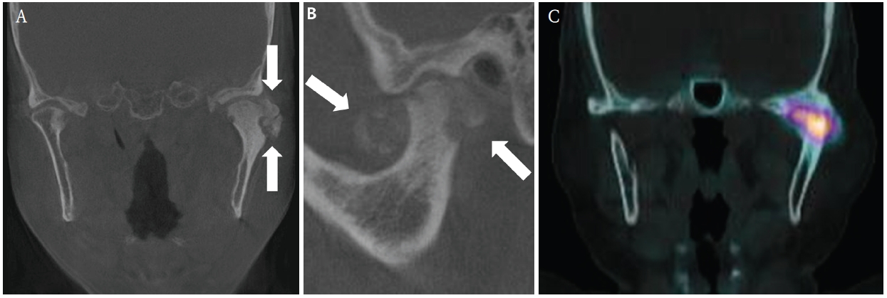
Radiographic findings of the left temporomandibular joint. A. Coronal cone-beam computed tomography (CBCT) image shows multiple calcified loose bodies (arrows) around the left condylar head. B. Sagittal CBCT image demonstrates bony erosion and sclerotic changes of the left condyle, along with multiple calcified loose bodies (arrows). C. Bone singlephoton emission computed tomography/computed tomography reveals increased radiotracer uptake in the left condyle, suggesting active bone metabolism.
MRI demonstrated a heterogeneous lesion involving the left TMJ with poorly defined and indistinct margins. The lesion involved the condylar head and was associated with cortical destruction and deformity. The internal architecture appeared irregular, without clear demarcation from surrounding structures, suggesting a possibly infiltrative growth pattern. These imaging features, particularly the ill-defined margins and osseous destruction, raised suspicion for a malignant tumor rather than a benign synovial condition (Fig. 2).
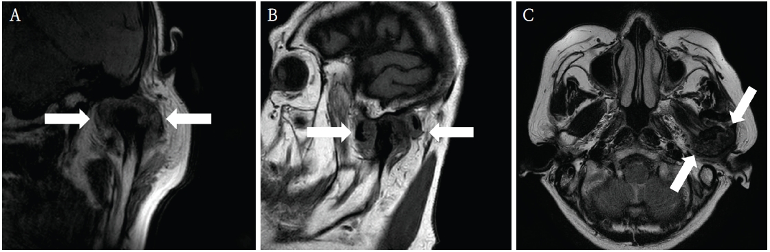
Magnetic resonance imaging findings of the left temporomandibular joint. A. Coronal view demonstrates a heterogeneous mass lesion (arrows) involving the left temporomandibular joint with poorly defined margins. B. Sagittal view shows the lesion (arrows) causing cortical destruction of the condylar head and irregular contour deformity. C. Axial view illustrates the extent of the lesion (arrows) around the temporomandibular joint with indistinct borders, suggesting an infiltrative growth pattern.
Despite several imaging features suggestive of a malignant tumor, including poorly defined margins and cortical destruction, the initial clinical impression favored synovial chondromatosis. This was primarily based on the presence of multiple calcified loose bodies within the joint space, a characteristic finding of synovial chondromatosis, along with associated sclerotic changes. In addition, the patient’s clinical presentation, including preauricular pain, swelling, and limited mouth opening, was consistent with a benign TMJ disorder. Based on this preliminary diagnosis, surgical intervention was planned under the assumption of a benign condition, with the aim of improving mandibular function and relieving symptoms. A preauricular approach was used to access the TMJ under general anesthesia. Intraoperatively, multiple creamy-white and brownish tissue fragments were identified within the joint space (Fig. 3). These materials appeared irregular in shape and were loosely distributed around the condylar head, initially suggesting cartilaginous loose bodies. Curettage was performed around the condylar head, including the surrounding joint space, followed by condyloplasty to improve joint contour. The articular disc, which was found to be medially displaced and folded, was repositioned. Postoperatively, the maximum mouth opening improved to 40 mm with satisfactory mandibular movement.
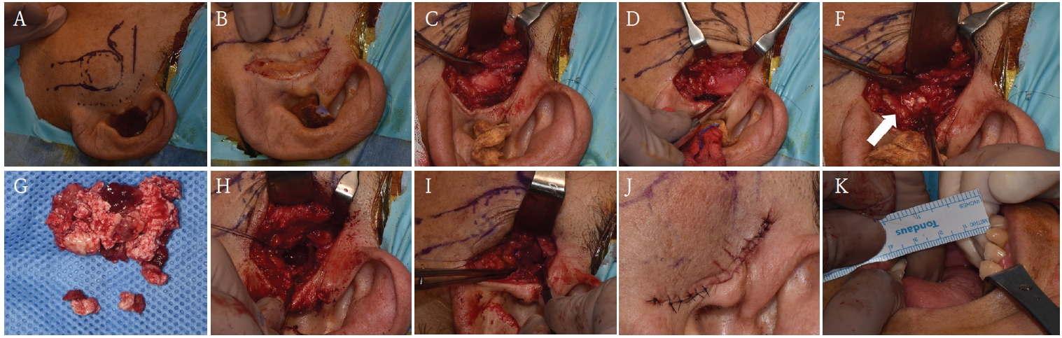
Intraoperative findings and surgical procedure of the left temporomandibular joint. A-D. Surgical exposure of the temporomandibular joint through a preauricular approach. E. Intra-articular view shows multiple creamy-white and brownish tissue fragments (arrows) within the joint space. F. Removed intra-articular tissue fragments, appearing as creamy-white and brownish masses. G. Intra-articular view after removal of the tissue, shows a cleared joint space around the condylar head. H. Condyloplasty with repositioning of the articular disc. I. Closure of the surgical site. J. Postoperative maximum mouth opening demonstrating improvement to approximately 40 mm.
Histopathological examination revealed grade III chondrosarcoma, characterized by high cellularity, nuclear pleomorphism, abundant mitoses, and disrupted lobular architecture. The histopathologic findings are consistent with grade III chondrosarcoma, with a Ki-67 proliferation index of approximately 10%. The final diagnosis was high-grade chondrosarcoma of the TMJ (Fig. 4). Given the high-grade diagnosis, additional surgical resection and adjuvant radiotherapy were recommended. However, the patient declined further treatment, citing improved function and age-related considerations. At the 12-month telephone follow-up, the patient remained asymptomatic and functionally stable, with no clinical evidence of disease progression.
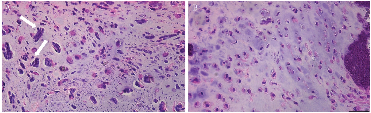
Histopathological findings of the temporomandibular joint lesion (H&E stain). A. High-power photomicrograph demonstrates hypercellularity, prominent nuclear pleomorphism, and multinucleated cells. The white arrows indicate cells with severe nuclear atypia and increased mitotic activity (original magnification, x400). B. Low-power photomicrograph showing the disrupted lobular architecture. These overall histopathological features are consistent with grade III chondrosarcoma (original magnification, x200).
Discussion
This case highlights the inherent diagnostic difficulty in distinguishing synovial chondromatosis from chondrosarcoma of the TMJ, especially given the rarity of both entities in this anatomic location. The patient's initial presentation, including preauricular pain, limited mandibular range of motion (comfortable mouth opening: 15 mm, maximum mouth opening: 25 mm), and imaging findings of condylar sclerosis, bony erosion, and calcified loose bodies, were all suggestive of synovial chondromatosis. However, the MRI findings of a poorly marginated mass and osseous destruction warranted further oncologic consideration.
Recent studies have proposed imaging-based criteria to improve diagnostic precision [6]. Among several evaluated radiologic markers, infiltration of the lateral pterygoid muscle tendon demonstrated the highest diagnostic value, followed by lesion size (typically > 4 cm), internal enhancement, and sclerosis of adjacent bone structures. If at least one of these four features is present, clinicians should include chondrosarcoma in the differential diagnosis. In fact, chondrosarcoma lesions tend to be larger in volume, with average dimensions around 3.9 cm × 1.7 cm, and frequently exceed 4 cm in maximum diameter [7,8].
Nevertheless, the overlapping clinical and radiologic features of synovial chondromatosis and chondrosarcoma often render preoperative differentiation elusive. Both entities can manifest with pain, trismus, condylar irregularity, and calcified intra-articular bodies. As such, diagnosis cannot rely solely on imaging [9]. In our case, radiologic findings such as multiple calcified loose bodies and sclerotic changes closely mimicked synovial chondromatosis, leading to an initial misdiagnosis. Notably, the final diagnosis of high-grade chondrosarcoma was established only after histopathologic examination, despite relatively benign-appearing imaging findings. This highlights the limitation of imaging alone and emphasizes the importance of histopathologic confirmation in ambiguous TMJ lesions.
However, histologic examination also has its limitations. The grading of chondrosarcoma is inherently subjective and often plagued by interobserver variability, especially in borderline or low-grade lesions [10]. Additionally, when chondrosarcoma arises in unconventional locations such as the TMJ, it may mimic the benign histologic architecture of synovial chondromatosis, further complicating diagnosis [11]. Although advanced imaging modalities such as MRI and PET-CT provide valuable insights into tumor biology and invasiveness, they remain insufficient for definitive diagnosis. Accordingly, a multidisciplinary approach that integrates clinical, radiological, and histopathological data is imperative. In select cases, intraoperative frozen section biopsy may serve as a pragmatic adjunct to guide real-time surgical decision-making, particularly in elderly patients or those presenting with high-risk imaging features such as cortical destruction or ill-defined margins.
Furthermore, this case underscores the ethical and therapeutic challenges posed by treatment refusal in geriatric oncology. Despite the histological confirmation of high-grade chondrosarcoma, the patient's decision to decline adjuvant therapy—coupled with her satisfactory functional status and lack of clinical recurrence at one year—raises the possibility that conservative surgery may be considered in exceptional cases with patient-centered goals of care. Nonetheless, long-term surveillance remains essential given the risk of local recurrence and distant metastasis associated with high-grade lesions [11].
In conclusion, accurately distinguishing synovial chondromatosis from chondrosarcoma of the TMJ is essential, given their markedly different biological behaviors and treatment strategies. This case highlights the limitations of relying solely on clinical and radiologic findings and underscores the importance of thorough histopathologic evaluation. Clinicians should maintain a high index of suspicion for malignancy in TMJ lesions presenting with atypical or aggressive features, even when initial impressions suggest a benign condition.
Notes
Conflicts of Interest
None