Clinical characteristics and surgical outcomes of lower lip mucocele: A retrospective study
Article information

Abstract
Purpose
This retrospective study investigated the clinical characteristics and surgical outcomes of lower lip mucocele at a single institution, focusing on the effectiveness of concomitant minor salivary gland removal in preventing recurrence.
Materials and Methods
Medical records of 19 patients who underwent surgery for lower lip mucocele at Inje University Busan Paik Hospital between 2011 and 2025 were retrospectively reviewed. Patients were classified into two groups: 1) Group A (n = 16) achieved complete resolution with a single procedure including concomitant excision of the mucocele and the underlying minor salivary gland(s) and 2) Group B (n = 3) experienced recurrence after primary excision at our institution and subsequently underwent re-operation with additional minor salivary gland removal. Among the 19 patients, 4 (Cases 1, 6, 8, and 9) had previously been treated at outside facilities before referral.
Results
The cohort comprised 12 males and 7 females, with a mean age of 22.1 years (range: 8–59 years). All 16 patients in Group A achieved resolution with a single operation. In Group B, recurrence developed within 2–5 weeks after primary excision; however, re-operation with additional minor salivary gland removal achieved resolution in all 3 patients. One patient developed transient commissure hypoesthesia, which partially recovered at final follow-up.
Conclusion
Concomitant excision of the mucocele and the underlying minor salivary gland(s) is effective in preventing recurrence. When recurrence occurs, additional gland removal achieves resolution. Minor salivary gland removal should be incorporated as a standard component of the surgical protocol for lower lip mucocele.
Introduction
Mucocele is a benign cystic lesion of the oral cavity that arises from mucus extravasation due to trauma to the duct of a minor salivary gland or from mucus retention caused by ductal obstruction. It is one of the most common soft tissue cystic lesions of the oral cavity, and the lower lip accounts for approximately 70–80% of all intraoral mucoceles, making it the most frequently affected site [1-3]. Trauma-induced ductal injury of the minor salivary glands is regarded as the predominant pathogenic mechanism [4,5], and a relatively higher incidence is observed in children and adolescents [6].
Clinically, lower lip mucocele presents as a fluctuant cystic swelling that is translucent, bluish, or normal in color, and tends to rupture and recur repeatedly [7,8]. Surgical excision is the standard treatment [9], and various alternative modalities have been reported, including CO2 laser ablation [10], marsupialization [11], and diode laser therapy [12].
Recurrence is the most significant clinical challenge in the management of oral mucocele, with reported recurrence rates ranging from 4% to 18% [8,13]. Incomplete removal of the surrounding minor salivary glands and repeated trauma are the primary causes of recurrence [4,13]. If a damaged ductal minor salivary gland remains in situ, mucus extravasation may recur. Therefore, concomitant removal of the minor salivary glands beneath and around the mucocele is expected to be effective in preventing recurrence; however, systematic analyses of this strategy remain limited [14,15].
This study retrospectively analyzed the medical records of patients who underwent surgery for lower lip mucocele at a single institution to evaluate the clinical effectiveness of combining mucocele excision with concomitant minor salivary gland removal, and to provide an evidence base for the optimal surgical strategy.
Materials and Methods
The medical records of patients who underwent surgery for lower lip mucocele at the Department of Oral and Maxillofacial Surgery, Inje University Busan Paik Hospital, between January 2011 and December 2025 were retrospectively reviewed. The study was conducted with waiver of informed consent under the approval of the Busan Paik Hospital Institutional Review Board (BPIRB 2026-03-042).
Inclusion criteria were patients diagnosed with lower lip mucocele who had undergone surgical excision at our institution and whose medical records contained sufficient clinical information. Exclusion criteria were: 1) final histopathological diagnosis of another cystic lesion; 2) lesion located at a site other than the lower lip (e.g., buccal mucosa, oral vestibule); and 3) patients who did not undergo surgery. Of the 21 patients initially reviewed, 2 were excluded according to these criteria, and a total of 19 patients were included in the final analysis. The diagnosis of lower lip mucocele was based on a combination of clinical and histopathological findings. Clinically, the diagnosis relied on the characteristic presentation of a fluctuant, translucent-to-bluish cystic swelling with a history of recurrent rupture, and was confirmed histopathologically in all cases.
Patients were classified into two groups based on the number of surgical procedures performed at our institution and the clinical course.
Group A (resolved with a single operation at our institution, n = 16): patients who achieved complete resolution with a single surgical procedure at our institution, which included concomitant excision of the mucocele and the underlying minor salivary gland(s). Among these, 2 patients (Cases 6 and 8) had previously been treated at outside facilities a local dental clinic (LDC) or an otolaryngology clinic and were referred to our institution following recurrence.
Group B (recurrence after primary excision at our institution → resolved after re-operation, n = 3): Patients who experienced recurrence after primary excision at our institution and subsequently underwent re-operation with additional minor salivary gland removal, achieving final resolution. Case 1 had previously undergone excision twice at an LDC before referral; recurrence was observed approximately 5 weeks after the primary operation at our institution, and a second operation with removal of 2 additional minor salivary glands (2 pieces) achieved resolution. Case 9 had a history of incision and drainage at an LDC before referral; recurrence occurred approximately 2 weeks after the primary operation at our institution, and a second operation with removal of minor salivary glands achieved resolution. Case 11 was treated exclusively at our institution; recurrence developed approximately 2 weeks after the primary excision, and a second operation (re-excision with additional minor salivary gland removal) achieved resolution.
All operations at our institution were performed under local infiltration anesthesia with 2% lidocaine and 1:100,000 epinephrine, in an outpatient, day surgery room (DSR), or inpatient setting. The standard surgical principle was to remove the minor salivary gland(s) directly beneath the mucocele simultaneously with excision of the mucocele. In Group B cases that experienced recurrence after the primary operation, re-operation included excision of the recurrent lesion and additional removal of the surrounding minor salivary glands. All excised specimens were submitted for histopathological examination, and wound closure was performed with absorbable sutures (Vicryl).
Collected data were analyzed using descriptive statistics. Continuous variables are expressed as mean ± standard deviation, and categorical variables are expressed as frequency and percentage.
Results
The final study population comprised 19 patients: 12 males (63.2%) and 7 females (36.8%). Age at first visit ranged from 8 to 59 years, with a mean of 22.1 ± 13.8 years. Children and adolescents (younger than 18 years) accounted for 6 patients (31.6%). Duration of symptoms ranged from 2 weeks to 9 months, and a history of trauma (e.g., lip biting) was identified in 6 patients (31.6%). Lesion size ranged from 3 mm to 14 mm (mean: 7.1 mm). The location was the right lower lip in 8 cases, the left lower lip in 9 cases, and the lower lip in 2 cases (Table 1). The mean follow-up duration was 29.1 ± 28.2 days (mean ± standard deviation). Clinical photograph showed a suspected mucocele on the right lower lip mucosa in Case 8 (Fig. 1).
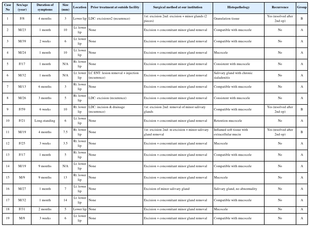
Patient demographics, lesion characteristics, surgical approach, and outcomes
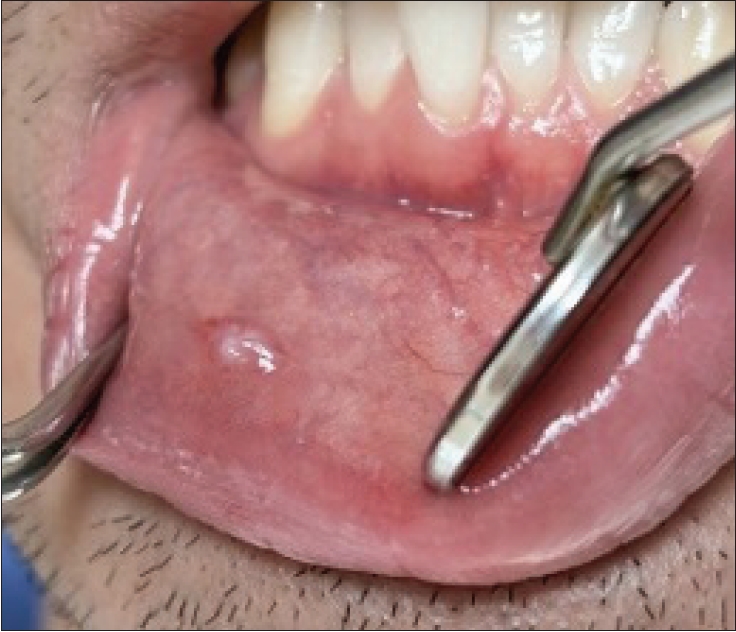
Clinical photograph shows a suspected mucocele on the right lower lip mucosa.
Of the 19 patients, 4 (Cases 1, 6, 8, and 9) had a history of treatment at outside facilities before referral to our institution. Case 1 had undergone excision twice at an LDC before presenting with recurrence; Case 6 had received lesion removal and injection therapy at an ENT clinic before recurrence; Case 8 had undergone excision at an LDC before recurrence; and Case 9 had undergone incision and drainage at an LDC before recurrence. The surgical methods used at the outside facilities were presumed to be simple excision or incision and drainage based on the medical records, and whether minor salivary gland removal had been performed could not be confirmed.
All 16 patients in Group A achieved complete resolution with a single surgical procedure at our institution, which included concomitant excision of the mucocele and the underlying minor salivary gland(s). No recurrence was observed during the follow-up period. Clinical photograph showed complete resolution of the mucocele lesion approximately 4 months after surgery in Case 8 (Fig. 2). Among these, Cases 6 and 8 had been referred following recurrence at outside facilities but achieved resolution with a single operation at our institution. The care setting was outpatient in 13 cases, day surgery room (DSR) in 5 cases, and inpatient in 1 case.
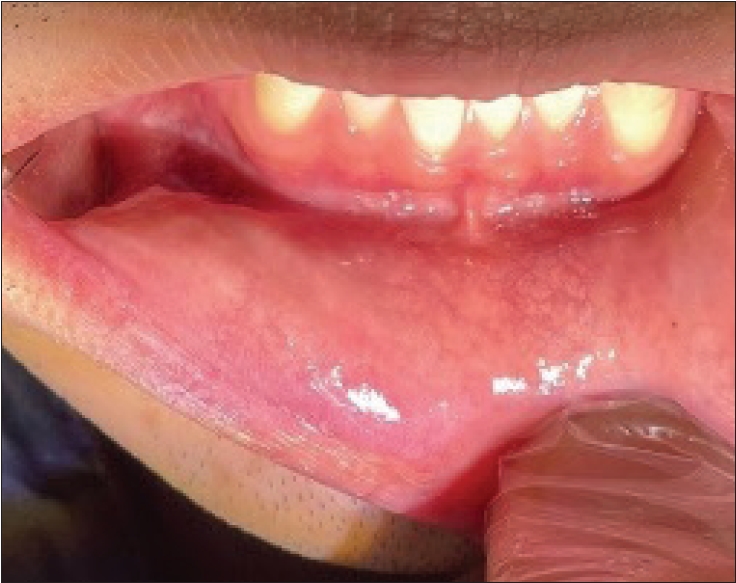
Clinical photograph shows complete resolution of the mucocele lesion approximately 4 months after surgery.
In Group B, Cases 1 and 9 had been referred from outside facilities following recurrence, whereas Case 11 had received treatment exclusively at our institution. Case 1 had undergone two excisions at an LDC before referral and developed recurrence approximately 5 weeks after the primary operation at our institution. A second operation with excision of the recurrent lesion and removal of 2 additional minor salivary glands (2 pieces) achieved resolution without further recurrence. Case 9 had a history of incision and drainage at an LDC before referral and developed recurrence approximately 2 weeks after the primary operation at our institution. A second operation with removal of minor salivary glands achieved resolution without further recurrence. Case 11 was treated exclusively at our institution and developed recurrence approximately 2 weeks after the primary excision. A second operation (re-excision with additional minor salivary gland removal) achieved resolution without further recurrence.
The surgical outcomes of all 19 patients are summarized in Table 2. All patients in Group A (n = 16) achieved complete resolution with a single operation, and all patients in Group B (n = 3) achieved final resolution after reoperation with additional minor salivary gland removal. The overall resolution rate was 100% in both groups following treatment at our institution.

Comparison of surgical outcomes between Group A and Group B
Histopathological results were available for all 19 cases (Table 3). Of these, 15 (78.9%) were confirmed as mucocele (extravasation or retention type) or compatible with mucocele. Additional findings included 1 case of granulation tissue, 1 of inflamed soft tissue with extracellular mucin, 1 of salivary gland with chronic sialadenitis, and 1 of salivary gland with no diagnostic abnormalities.
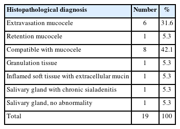
Histopathological findings
One patient in Group A (Case 17) developed hypoesthesia of the left commissure region (30–40% sensation) beginning on the day after surgery. This was attributed to a transient neuropraxia of a branch of the mental nerve related to the surgical manipulation. At the final follow-up (approximately 2 months postoperatively), sensation had partially recovered to 50%. No other complications, including hemorrhage, infection, or wound dehiscence, were observed.
Discussion
Lower lip mucocele is one of the most common benign cystic lesions of the oral cavity, with mucus extravasation caused by traumatic ductal injury of the minor salivary glands as the predominant pathogenic mechanism [1-5]. This study retrospectively analyzed 19 patients who underwent surgery for lower lip mucocele at a single institution, evaluating the clinical effectiveness of concomitant minor salivary gland removal combined with mucocele excision.
The mean age in the present study was 22.1 years, with children and adolescents accounting for approximately 32% of the cohort, consistent with previous reports [2,6]. Wu et al. [6] reported that lower lip mucocele in pediatric patients may be associated with a higher recurrence rate compared with adults, potentially related to the higher frequency of repetitive lip trauma in this age group. Of the 19 cases with available histopathological results, 15 (78.9%) were confirmed as mucocele or compatible findings, indicating relatively high accuracy of clinical diagnosis.
The most important finding of this study was the high resolution rate achieved when surgery included concomitant minor salivary gland removal. All 16 patients in Group A achieved complete resolution with a single operation, with no recurrence during the follow-up period. Notably, Cases 6 and 8, who had been referred to our institution following recurrence at outside facilities, also achieved resolution with a single operation at our institution. This outcome underscores the clinical significance of concomitant minor salivary gland removal.
In Group B, 3 patients (Cases 1, 9, and 11) experienced recurrence after the primary excision at our institution. Case 1 had undergone two excisions at an LDC before referral and developed recurrence approximately 5 weeks postoperatively. Case 9 had a history of incision and drainage at an LDC before referral and developed recurrence approximately 2 weeks postoperatively. Case 11, treated exclusively at our institution, experienced recurrence approximately 2 weeks after the primary excision. Possible contributing factors to recurrence in these cases include fibrosis of the perifocal tissue from repeated trauma and surgery, and involvement of multiple minor salivary glands. After re-operation with sufficient additional removal of the surrounding minor salivary glands, all three cases achieved resolution without further recurrence.
Understanding the anatomical characteristics of the minor salivary glands is essential for comprehending the mechanism of mucocele recurrence. Hundreds of minor salivary glands are densely distributed beneath the lower lip mucosa, and their ducts are susceptible to repeated traumatic injury [4,14]. Even if the cystic lesion alone is removed, persistence of a minor salivary gland with a damaged duct may lead to recurrent mucus extravasation. Bowers and Schaitkin [15] emphasized that complete removal of the causative minor salivary gland is central to mucocele treatment, and that wide excision including adjacent glands is necessary for recurrent lesions. Choi et al. [13] also reported that whether minor salivary gland removal was performed was a significant predictor of recurrence. The results of this study support these previous findings, and the achievement of complete resolution in all 16 Group A patients with a single operation through concomitant gland removal directly supports the clinical utility of this approach.
Regarding the surgical strategy for recurrent cases (Group B), this study confirmed that additional removal of the surrounding minor salivary glands in conjunction with excision of the recurrent lesion is effective when recurrence occurs after simple excision. In all three Group B cases, re-operation with additional gland removal achieved resolution. This demonstrates that complete resolution can be achieved through re-operation including gland removal even in recurrent cases, and suggests that minor salivary gland removal is a key component of both primary and revision surgery.
Regarding complications, 1 case developed postoperative hypoesthesia of a branch of the mental nerve, which partially recovered. Because the minor salivary glands of the lower lip are densely distributed around the mental foramen, careful anatomical awareness and precise surgical technique are required to avoid injury to mental nerve branches during gland removal [9].
Limitations of this study include the retrospective design, small sample size, heterogeneity of follow-up duration, and inability to confirm the precise surgical methods used at outside facilities. In addition, the mean follow-up duration in this study was relatively short (29.1 ± 28.2 days), and follow-up time varied considerably among patients. Because recurrence in Group B occurred within 2–5 weeks of the primary operation, a subset of patients—particularly those with shorter follow-up—may not have been observed long enough to reliably exclude late recurrence. Longer and more uniform follow-up would be needed to more accurately determine the true recurrence rate. Prospective studies with larger cohorts and longer follow-up periods are warranted to validate the findings of this study.
In conclusion, this single-institution retrospective study of 19 patients with lower lip mucocele confirmed that surgical excision combined with concomitant minor salivary gland removal is an effective approach for preventing recurrence and achieving resolution.
Notes
Conflicts of Interest
None
Acknowledgement
The author acknowledges the use of generative AI for drafting and grammatical refinement. All AI-generated suggestions and conclusions were critically reviewed and verified by the author, who remains responsible for the integrity of the work.