Autotransplantation of a mature third molar into a graft-free maxillary sinus: A 9-year follow-up case report
Article information

Abstract
This report describes periodontal ligament (PDL)–preserving autotransplantation combined with graft-free crestal sinus membrane elevation in a posterior maxillary site with limited residual bone height. A 28-year-old woman with a non-restorable maxillary first molar and approximately 3 mm of residual crestal bone height underwent autotransplantation of a mature third molar with simultaneous graft-free sinus membrane elevation. Approximately nine years of follow-up demonstrated stable function with preservation of the PDL space, no ankylosis or pathologic root resorption, and progressive bone formation within the sinus-associated void. PDL-preserving autotransplantation with graft-free sinus membrane elevation may represent a viable biologically based alternative in selected posterior maxillary cases.
Introduction
Tooth autotransplantation can yield favorable outcomes when appropriate case selection and surgical principles are respected [1]. However, its clinical use has declined with the widespread adoption of dental implant therapy, driven by technical sensitivity, limited recipient-site bone volume, the frequent need for endodontic treatment in mature donor teeth, and the predictability of contemporary implant systems.
From a biologic perspective, autotransplantation uniquely preserves the periodontal ligament (PDL), which provides shock absorption, proprioception, and cellular sources for tissue homeostasis and repair [2–4]. In contrast, implant-based replacement lacks a PDL and connective tissue attachment, and peri-implant soft tissues demonstrate a different barrier configuration that may be more susceptible to microbial challenge than the periodontal attachment apparatus [5–7].
A major limitation to broader application of autotransplantation is insufficient vertical bone height, particularly in the posterior maxilla where sinus pneumatization may necessitate sinus floor elevation. While sinus membrane elevation with or without grafting is well established for implant therapy, integration with tooth autotransplantation—especially in graft-free settings—has been infrequently reported, and long-term evidence remains limited [8–10]. This report describes a PDL-preserving autotransplantation combined with graft-free crestal sinus membrane elevation with nine-year clinical and radiographic follow-up.
Case Report
This study was exempted from Institutional Review Board (IRB) review by a government-designated public Institutional Bioethics Committee (Exemption No. P01-202603-01-012). The study was conducted in accordance with the Declaration of Helsinki. Written informed consent was obtained from the patient for clinical treatment and for publication of anonymized clinical and radiographic data.
A 28-year-old woman presented at the initial visit (Day 0) with a chief complaint related to the maxillary left first molar (tooth 26), which had advanced root caries and complete coronal destruction. Clinical examination revealed bilateral absence of mandibular first molars and the presence of all four third molars. The maxillary right third molar was mesially inclined and slightly undererupted; the maxillary left third molar was fully impacted with mesial angulation. Both mandibular third molars were fully erupted with partial distal gingival coverage. Preoperative panoramic and cone-beam computed tomography (CBCT) findings are shown in Figures 1 and 2.
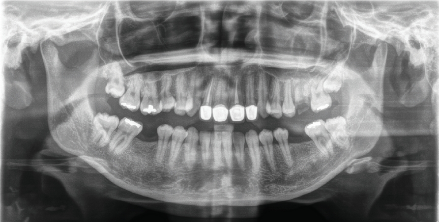
Preoperative panoramic radiograph obtained at the initial visit. The image shows advanced destruction of the maxillary left first molar (tooth 26), bilateral absence of the mandibular first molars, and the presence of all four third molars, which are later evaluated as potential donor teeth.
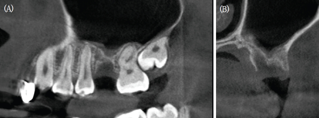
CBCT images of the maxillary left first molar recipient site obtained 1 month after extraction of tooth 26. Sagittal (A) and buccopalatal (B) views demonstrate adequate mesiodistal and buccopalatal dimensions at the cervical aspect of the alveolar ridge with limited residual vertical bone height. Crestal bone height from the alveolar crest to the sinus floor measures approximately 3 mm at the central portion of the site.
The patient preferred replacement using her own teeth rather than implant-supported restorations. Autotransplantation was not feasible in mandibular molar sites because of insufficient ridge width; implant therapy was planned for those areas. The maxillary left first molar site was considered a potential recipient site after extraction, although limited vertical bone height was anticipated.
Tooth 26 was extracted 4 days after the initial visit. CBCT obtained approximately 5 weeks after the initial visit demonstrated adequate mesiodistal (7.17 mm) and buccopalatal (10.6 mm) dimensions at the cervical aspect of the alveolar ridge. Residual crestal bone height between the alveolar crest and maxillary sinus floor was approximately 3 mm, which was insufficient for conventional autotransplantation without sinus intervention.
The patient was informed that autotransplantation would require simultaneous sinus membrane elevation. After discussion of alternatives, including implant placement with sinus grafting, the patient consented to autotransplantation combined with graft-free crestal sinus elevation. The maxillary right third molar was selected as the donor tooth based on root morphology and size compatibility (maximum cervical dimensions approximately 8.0 × 9.0 mm; total root length 9 mm).
Autotransplantation was performed approximately 5 weeks after the initial visit under local anesthesia. The donor tooth was atraumatically extracted with meticulous preservation of the PDL. The recipient site was prepared, followed by crestal approach elevation of the Schneiderian membrane without lateral window access. No bone grafts or biomaterials were placed.
The donor tooth was positioned partially within the elevated sinus space, maintaining a graft-free compartment filled only with blood clot. Primary stability was achieved through socket adaptation, and the transplanted tooth was stabilized with sutures during the early healing phase. Immediate postoperative stabilization is shown in Figure 3.
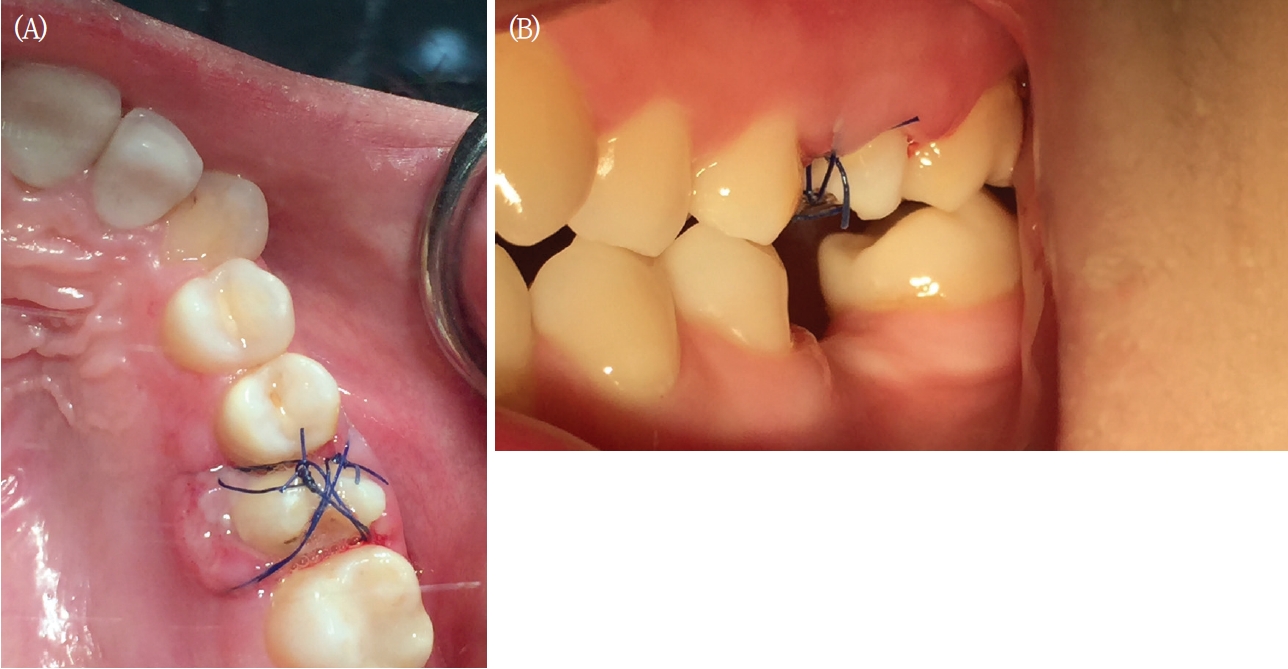
Immediate postoperative intraoral photographs show stabilization of the transplanted tooth using sutures after autotransplantation at the maxillary left first molar site. The tooth is positioned slightly subocclusal relative to adjacent teeth to ensure complete placement of the periodontal ligament-covered root within residual crestal bone and to minimize early functional loading.
To maximize preservation of PDL vitality, recipient-site preparation was completed before final extraction of the donor tooth, thereby minimizing extraoral time. Following extraction, the donor tooth was temporarily maintained within its original socket until transfer to the recipient site. Particular care was taken to perform atraumatic extraction to minimize damage to the root surface and periodontal ligament.
Pulp extirpation was performed approximately 2 weeks after the autotransplantation to reduce the risk of inflammatory complications associated with a mature donor tooth. A provisional crown was placed approximately 5 weeks after the autotransplantation to protect the transplanted tooth and allow controlled functional loading. Root canal treatment was completed approximately 7 months after the autotransplantation, followed by definitive prosthodontic restoration with a zirconia crown approximately 11 months after the autotransplantation. The completion of root canal treatment was delayed because the patient traveled overseas for an extended business-related stay during treatment. Surgical care was performed by an oral and maxillofacial surgeon, and endodontic treatment was performed by an endodontist.
The patient underwent periodic clinical and radiographic follow-up for approximately nine years. Periapical radiographs, panoramic radiographs, and CBCT imaging demonstrated preservation of a continuous PDL space, stable crestal bone levels, and no evidence of ankylosis or pathologic root resorption (Figs. 4 and 5).
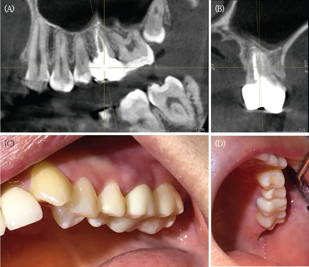
Longitudinal radiographic and clinical evaluation. CBCT images obtained 3 years after the autotransplantation show sagittal (A) and buccopalatal (B) views with a continuous periodontal ligament space and bone formation in the region corresponding to the prior sinus-associated void. Intraoral photographs obtained 4 years after the autotransplantation show buccal (C) and palatal (D) views demonstrating healthy peri-dental soft tissue architecture and absence of clinical inflammation.
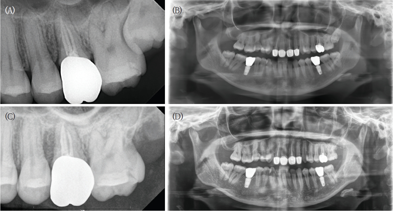
Long-term radiographic follow-up. Periapical (A) and panoramic (B) radiographs obtained 4 years and 6 months after the autotransplantation. Periapical (C) and panoramic (D) radiographs obtained 9 years after the autotransplantation. Radiographs demonstrate stable crestal bone levels and a well-defined PDL space without ankylosis, root resorption, or progressive bone loss.
Progressive radiographic bone formation developed around the transplanted root within the sinus cavity despite absence of graft material. Newly formed bone thickness surrounding the root was approximately 3–5 mm radiographically. At the most recent follow-up, nine years after the autotransplantation, mild buccal gingival recession was present and was interpreted as physiologic soft-tissue adaptation in the absence of inflammation, progressive bone loss, or loss of PDL continuity. The patient reported no pain or mobility, with only intermittent tenderness during systemic fatigue that resolved spontaneously.
Representative intraoral findings at the most recent visit are shown in Figure 6.
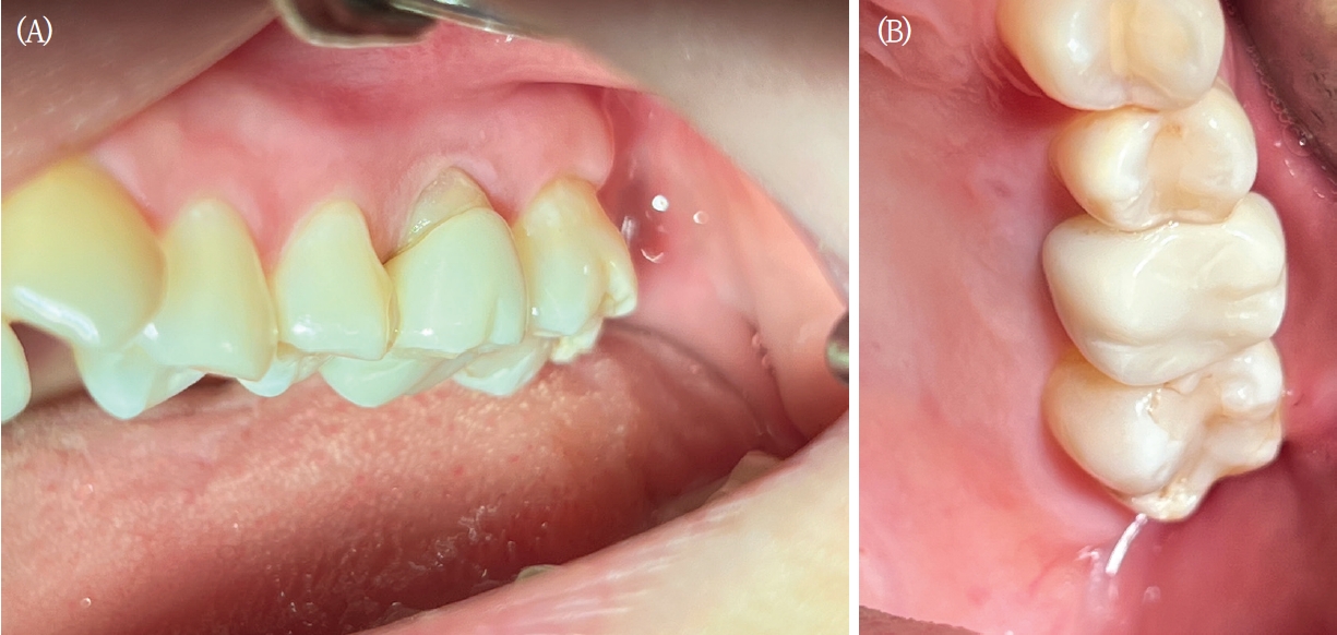
Most recent intraoral photographs obtained nine years after the autotransplantation. A. Buccal view shows mild gingival recession of approximately 2 mm without clinical inflammation. B. Palatal view shows stable gingival architecture comparable to earlier follow-up images.
Discussion
This case demonstrates long-term clinical and radiographic stability after PDL-preserving autotransplantation combined with graft-free crestal sinus membrane elevation in a posterior maxillary site with limited residual bone height. The nine-year outcome with a maintained PDL space and absence of ankylosis or pathologic root resorption supports the biologic plausibility that PDL preservation contributes to periodontal adaptation and stability [2–4].
In the atrophic posterior maxilla, sinus membrane elevation is commonly performed with grafts or biomaterials, particularly in implant therapy [8–9]. In the present approach, grafting was intentionally avoided to preserve native healing processes and to reduce concern that particulate graft materials in close proximity to the donor root might interfere with PDL-mediated healing or increase the risk of ankylosis [1,10]. Although histologic confirmation is not feasible in a clinical case report, long-term maintenance of a continuous PDL space with no ankylosis or pathologic resorption provides clinically meaningful indirect evidence of periodontal stability [1].
Mild buccal gingival recession was observed at long-term follow-up. A biologically plausible explanation is that a localized soft-tissue interface developed against enamel rather than cementum. This may occur when a donor tooth is positioned slightly subcrestally to ensure full embedding of cementum-covered root within available bone, while recipient-site morphology cannot precisely reproduce the cemento-enamel junction contour. In such areas, connective tissue attachment with Sharpey fiber insertion is not expected, and the seal may rely predominantly on junctional epithelial attachment [2,5]. Compared with connective tissue attachment to cementum, this configuration may be less resistant over time under functional loading and physiologic remodeling, potentially predisposing to site-specific recession. Stability on the palatal aspect supports an asymmetric, interface-dependent response [6–7].
In the present case, positioning of the donor tooth was guided by the objective of ensuring that the entire PDL-covered root surface remained within the available alveolar bone. Because the cemento-enamel junction was not circumferentially uniform, placement based on the most coronal aspect of the CEJ resulted in slight subcrestal positioning in some areas. In addition, because simultaneous sinus membrane elevation reduced the need for extensive recipient-site bone preparation, the potential benefit of a replica tooth for repeated socket adjustment was considered limited.
This report is limited by its single-case design and lack of histologic confirmation; therefore, generalization is not possible. Nevertheless, the extended follow-up suggests that graft-free sinus membrane elevation can be combined with PDL-preserving autotransplantation in carefully selected posterior maxillary cases.
Although several reports have described autotransplantation combined with sinus floor elevation, the present case is distinguished by the use of a mature third molar donor tooth, the complete absence of grafting materials, and approximately nine years of clinical and radiographic follow-up. These features provide unique long-term evidence supporting the biologic regenerative potential of the periodontal ligament in sinus-associated defects.
The present case also suggests that maintenance of a viable periodontal ligament may contribute not only to tooth retention but also to bone regeneration within an elevated sinus environment.
PDL-preserving autotransplantation combined with graft-free crestal sinus membrane elevation demonstrated sustained clinical function and radiographic stability over approximately nine years in an atrophic posterior maxillary site. This outcome supports further investigation of biologically grounded surgical strategies that leverage the regenerative potential of the PDL in sinus-associated defects.
Notes
Conflicts of Interest
None
Acknowledgement
The author used ChatGPT for language refinement and manuscript organization. The author reviewed and approved all content and takes full responsibility for the manuscript.