Angioleiomyoma of the hard palate: Report of a case and clinical, imaging, immunohistochemical analysis
-
Euijune Chang1
 , Donggi Ahn2
 , Jun-Young Kim1,*
- Received May 12, 2025 Revised May 27, 2025 Accepted June 4, 2025
- ABSTRACT
-
Angioleiomyoma, also known as vascular myoma or vascular leiomyoma, is a rare benign tumor originating from smooth muscle cells of the tunica media of blood vessels, characterized by numerous blood vessels and spindle-shaped smooth muscle cells. A 40-year-old Korean female was referred with a 14 × 8 × 6 mm pedunculated, elastic mass on the right hard palate. She reported the lesion had been present for one year without pain or noticeable growth. MRI showed a uniform signal pattern with heterogeneous hypo-intensity on T1-weighted and hyper-intensity on T2-weighted images. The tumor was excised under local anesthesia. Histopathology showed cytoplasmic positivity for SMA, Desmin, and Caldesmon, and negativity for pan-cytokeratin, S-100, CD31, CD34, and HMB45. Based on these findings, the lesion was diagnosed as angioleiomyoma. This represents the first reported case of angioleiomyoma occurring in the hard palate in Korea.
- Introduction
- Introduction
Leiomyoma, a benign tumor originating from smooth muscle tissue, is classified by the world health organization (WHO) into three histopathological subtypes; solid leiomyoma, vascular leiomyoma (angiomyoma, angioleiomyoma), and epithelioid leiomyoma [1]. Angioleiomyomas arise from the smooth muscle cells of the tunica media and are commonly found in the uterine myometrium, gastrointestinal tract, and lower limbs of middle-aged individuals. However, these tumors are rarely found in the oral cavity due to the limited presence of smooth muscle in the head and neck region [2]. Leiomyomas in the oral cavity account for less than 1% of all cases, with oral leiomyomas comprising only 0.06% of all leiomyomas [3]. We report a case of angioleiomyoma in the hard palate, which is classified as a vascular leiomyoma, a benign smooth muscle tumor with prominent vascular components, highlighting its MRI features and histopathological characteristics.
- Case Report
- Case Report
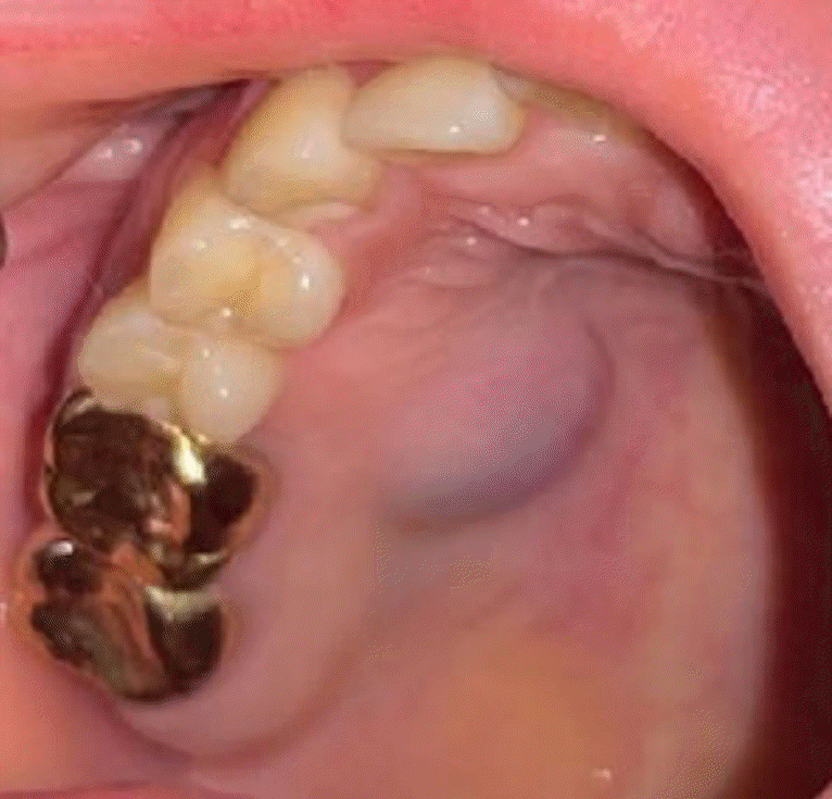
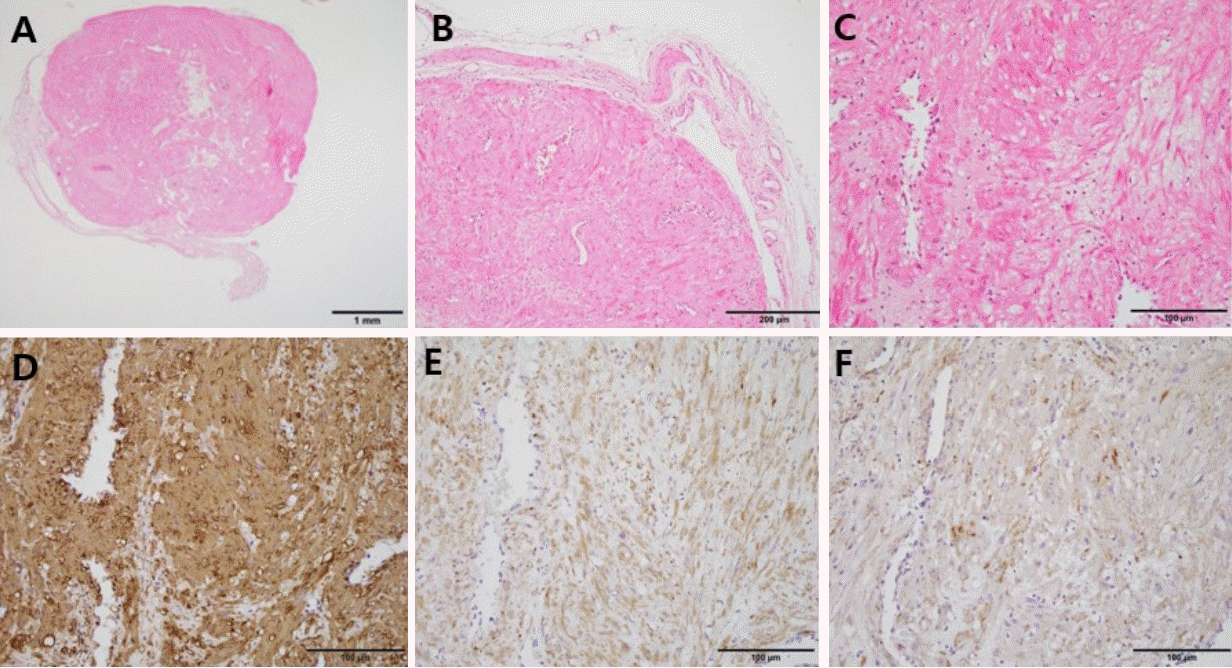
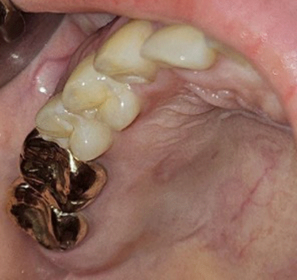
This study was approved by the institutional review board (IRB) of yonsei university dental hospital (IRB No.2-2025-0017). Informed consent was not required for the case report as no patient-specific information was disclosed.A 40-year-old Korean female patient was referred to our hospital for a tumor located in right hard palate that had been present for a year with no significant progression in size (Fig. 1). The lesion had no associated features, and the patient did not recall any preceding events that might have precipitated the swelling. There were no accompanying dental symptoms. The patient had no significant medical history and was not taking any regular medications. Clinical examination revealed a pedunculated, round, elastic, soft, red-purple mass, approximately 14 mm x 8 mm x 6 mm in size overlying with normal mucosa. The mass was nontender to palpation. A diagnostic magnetic resonance imaging (MRI) examination was obtained to determine the margin of the mass and assess for any intranasal or intra-antral involvement. MRI showed a well-circumscribed mass on the hard palate, uniform signal pattern with heterogeneous hypo-intensity on T1- weighted and hyper-intensity on T2-weighted and flow-like void inside the mass (Fig. 2). The mass was excised under local anesthesia and sent for histologic assessment.Histopathologically, this solitary lesion is encapsulated by thin to thick fibrous tissue (Fig. 3A and B). It consists of intertwining bundles of eosinophilic and spindle cells among numerous blood vessels. This tumor cells predominantly have oval nuclei, though some exhibited elongated, cigar-shaped nuclei with no evidence of nuclear atypia, including abnormal mitotic figure, nuclear pleomorphism, or necrosis (Fig. 3C). Immunohistochemically, the spindle cells show cytoplasmic positivity for smooth muscle actin (SMA), Desmin, and Caldesmon but are negative for pan-cytokeratin, S100, CD31, CD34 and HMB45(Fig. 3D-F). The Ki-67 labeling index is also very low, nearly 0% (<1%). The diagnosis of angioleiomyoma was confirmed by the characteristic histomorphological features, supported by the positive expression of smooth muscle markers. The patient returned for a follow-up visit two weeks postoperatively. Clinical examination confirmed that the palatal mucosa had healed well, with no signs of postoperative infection or recurrence of the tumor (Fig. 4).
- Discussion
- Discussion
Angioleiomyoma is a benign mesenchymal soft tissue tumor originating from vascular smooth muscle. Angioleiomyoma is rarely encountered in the oral cavity, as smooth muscle is primarily found in the tunica media of blood vessels and with the most commonly affected sites being the lips (48.6%), tongue (9.2%), hard palate (9.2%) and buccal mucosa (9.2%) [4]. This tumor typically presents as a slow-growing, painless, and well-circumscribed mass [2]. However, some cases report associated pain, which may be due to nerve involvement or vascular compression [5].MRI characteristics of oral angioleiomyomas are not well-documented due to their rarity, but as seen in this case, they typically present as well-defined masses with heterogeneous signal intensity patterns [3]. Recent studies highlight the importance of MRI in preoperative assessment, MRI features such as hyperintensity on T2-weighted imaging with hypointense rim corresponding to a fibrous capsule, isointense to slightly hyperintense compared to muscle on T1-weighted images and uniform enhancement on post-contrast images [6]. It is crucial to differentiate angioleiomyomas from other benign tumors that can develop in the palate (e.g. neurilemmomas, neurofibroma, pleomorphic adenoma) using MRI for accurate diagnosis and treatment planning in advance to the surgical intervention. Neurilemmomas have a very distinctive appearance with a target sign on peripheral nerves in MRI. Neurofibroma tend to show low-to-intermediate signal intensity on T1-weighted images and a heterogeneous appearances on T2-weighted images [7]. These imaging characteristics enable clinicians to exclude neurofibromas and neurilemmomas from the differential diagnosis. Pleomorphic adenomas present with imaging findings similar to angioleiomyomas, including a predilection for homogeneous intermediate signal intensity on T1-weighted images and heterogeneous high signal intensity on T2-weighted images [8]. Advanced imaging modalities such as CT scans and doppler ultrasonography may help in assessing the lesion’s vascular nature and detecting blood flow patterns within the tumor. However, due to overlapping features with other mesenchymal neoplasms, histopathological confirmation remains the gold standard. Histopathologically, angioleiomyomas is classified into three subtypes: solid, venous and cavernous. The solid-type consists of compacted bundles of smooth muscle cells and thinwalled blood vessels, while the venous-type exhibits thick-walled vascular channels that blend with smooth muscle bundles. The cavernous-type, in contrast, is characterized by large vascular channels with thin muscular walls [9]. The differential diagnosis of angioleiomyoma in the oral cavity is crucial due to its resemblance to other soft tissue neoplasm, including pyogenic granuloma, peripheral giant cell granuloma, peripheral ossifying fibroma, schwannoma, and minor salivary gland tumors [5]. Immunohistochemical analysis is essential for differential diagnosis, as angioleiomyomas stain positively for SMA, Desmin, and Caldesmon while lacking markers for epithelial, neural, and endothelial cells [10]. In this case, immunohistochemical staining for SMA, Desmin, Caldesmon, S100, CD31, CD34 and HMB45 were performed. SMA serves as the key immunomarker of smooth muscle cells, Desmin staining identifies a specific intermediate filament protein in muscle cell and Caldesmon, an actinbinding protein, regulates contraction in smooth muscle cells. Our case was confirmed as a muscle tumor, differentiated from other spindle cell tumors based on tests using these markers. Unlike these findings, all the other markers showed negative results. Negativity for S100 helps exclude the possibility of neural tumors such as schwannomas and neurofibromas, because this marker is expressed in schwann cells, melanocytes, and certain dendritic cells. in addition, vascular tumors such as hemangiomas and angiosarcomas were excluded from the final diagnosis due to negative immunohistochemical sensitivity to CD31, CD34 which are specific markers expressed in vascular endothelial cells. these tumor cells were also negative for HMB45 staining, the melanocytic differentiation marker, leading to exclusion of melanocytic tumors including spindle cell melanoma [11].Given their benign nature, complete surgical excision is the treatment of choice, with an excellent prognosis and low recurrence rates. Although rare, recurrence may occur, particularly in cases of incomplete excision [12].Genetic and molecular studies suggest potential roles of hormonal influences and genetic predispositions in the development of angioleiomyomas [13]. Further research should focus on the underlying pathophysiology, molecular characteristics, and potential genetic factors associated with oral angioleiomyomas to better understand their etiology and improve management strategies.
- NOTES
- NOTES
Fig. 1.
Intraoral clinical photograph (pre-operation) showing round, elastic, red-purple mass in the right side of the hard palate. (14 mm x 8 mm x 6 mm in size).

Fig. 2.
Pre-operation magnetic resonance images (MRI). A. Axial T1-weight image showing a well-circumscribed mass on the right side of the palate (red arrow). B and C. Axial, coronal T2-weighted image showing hyperintensity and flow-like void inside the mass (red arrows).

Fig. 3.
Histopathological findings. A and B. The lesion is encapsulated by fibrous tissue and predominantly composed of proliferative, spindle cells interspersed among numerous thick-walled blood vessels (A. H&E staining, x12.5, B. H&E staining, x100). C. These eosinophilic, spindle cells exhibit oval to elongated nuclei without any nuclear atypia and are arranged in interlacing bundles (H&E staining, x200). D-F. The tumor cells are cytoplasmic positive for smooth muscle markers (D. SMA, E. Desmin, and F. Caldesmon).

Fig. 4.
Intraoral clinical photograph (post-operation; 2 weeks) showing well palatal mucosa healing and coverage. No sign of post operative infection or recurrence of the tumor.

- REFERENCES
- REFERENCES
- 1. Arpağ OF, Damlar I, Kılıç S, Altan A, Taş ZA, Özgür T. Angioleiomyoma of the gingiva: a report of two cases. J Korean Assoc Oral Maxillofac Surg 2016;42:115–9.
[Article] [PubMed] [PMC]2. Eley KA, Alroyayamina S, Golding SJ, Tiam RN, Watt-Smith SR. Angioleiomyoma of the hard palate: report of a case and review of the literature and magnetic resonance imaging findings of this rare entity. Oral Surg Oral Med Oral Pathol Oral Radiol 2012;114:e45–9.
[Article] [PubMed]3. Matiakis A, Karakostas P, Pavlou AM, Anagnostou E, Poulopoulos A. Angioleiomyoma of the oral cavity: a case report and brief review of the literature. J Korean Assoc Oral Maxillofac Surg 2018;44:136–9.
[Article] [PubMed] [PMC]4. Brooks JK, Nikitakis NG, Goodman NJ, Levy BA. Clinicopathologic characterization of oral angioleiomyomas. Oral Surg Oral Med Oral Pathol Oral Radiol Endod 2002;94:221–7.
[Article] [PubMed]5. Liu Y, Li B, Li L, Liu Y, Wang C, Zha L. Angioleiomyomas in the head and neck: a retrospective clinical and immunohistochemical analysis. Oncol Lett 2014;8:241–7.
[Article] [PubMed] [PMC]6. Agarwal P, Creanga AD. Vascular leiomyoma of oral cavity: a case report in young male patient. J Oral Maxillofac Pathol 2024;28:506–10.
[Article] [PubMed] [PMC]7. Hillier JC, Moskovic E. The soft-tissue manifestations of neurofibromatosis type 1. Clin Radiol 2005;60:960–7.
[Article] [PubMed]8. Hisatomi M, Asaumi J, Yanagi Y, Konouchi H, Matsuzaki H, Honda Y et al. Assessment of pleomorphic adenomas using MRI and dynamic contrast enhanced MRI. Oral Oncol 2003;39:574–9.
[Article] [PubMed]9. Ramesh P, Annapureddy SR, Khan F, Sutaria PD. Angioleiom yoma: a clinical, pathological and radiological review. Int J Clin Pract 2004;58:587–91.
[Article] [PubMed]10. Tsuji T, Satoh K, Nakano H, Kogo M. Clinical characteristics of angioleiomyoma of the hard palate: report of a case and an analysis of the reported cases. J Oral Maxillofac Surg 2014;72:920–6.
[Article] [PubMed]11. Jordan RC, Regezi JA. Oral spindle cell neoplasms: a review of 307 cases. Oral Surg Oral Med Oral Pathol Oral Radiol Endod 2003;95:717–24.
[Article] [PubMed]
