


The use of buccal fat free graft in palatal defect after removal of palatal pleomorphic adenoma: A case report
-
Si-On Choi
 , Hoi-Bin Jeong, Jin-A Baek*
- Received March 27, 2025 Revised June 23, 2025 Accepted July 28, 2025
- ABSTRACT
-
Autologous buccal fat pad graft is widely utilized for the closure of oroantral and oronasal fistulas, as well as for reconstructing intraoral post-operative soft tissue defects. However, its primary use is as a pedicled flap, which may lead to complications such as necrosis, graft failure, hematoma, infection, facial nerve injury, arterial bleeding, donor site morbidity, trismus, wound dehiscence, and excessive granulomatous tissue formation. To address these limitations, numerous clinical reports have emerged advocating the use of buccal fat free grafts. In this manuscript, buccal fat free graft was performed on a 26-year-old female patient following the removal of a pleomorphic adenoma on the palatal side, resulting in successful healing. This illustrates that buccal fat free graft may be an effective surgical method for the reconstruction of various intraoral defects.
- Introduction
- Introduction
Autologous buccal fat pad graft is widely utilized for the closure of oroantral and oronasal fistulas, as well as for reconstructing intraoral post-operative soft tissue defects. The widespread use of the buccal fat pad graft is attributed to ease of harvesting, low complication rates, and a reputation as a relatively safe procedure with high success rate. However, its primary use is as a pedicled flap, which may lead to complications such as necrosis, graft failure, hematoma, infection, facial nerve injury, arterial bleeding, donor site morbidity, trismus, wound dehiscence, and excessive granulomatous tissue formation [1]. Among these limitations, the application of a pedicled flap is particularly challenging when the oral defect is located far from the buccal fat pad, as this may result in tissue contraction and functional impairments, such as restricted mouth opening. To address these limitations, numerous clinical reports have emerged advocating the use of buccal fat free grafts. In this manuscript, buccal fat free graft was performed on a 26-year-old female patient following the removal of a pleomorphic adenoma on the palatal side, resulting in successful healing. This illustrates that buccal fat free graft may be an effective surgical method for the reconstruction of various intraoral defects.
- Case Report
- Case Report
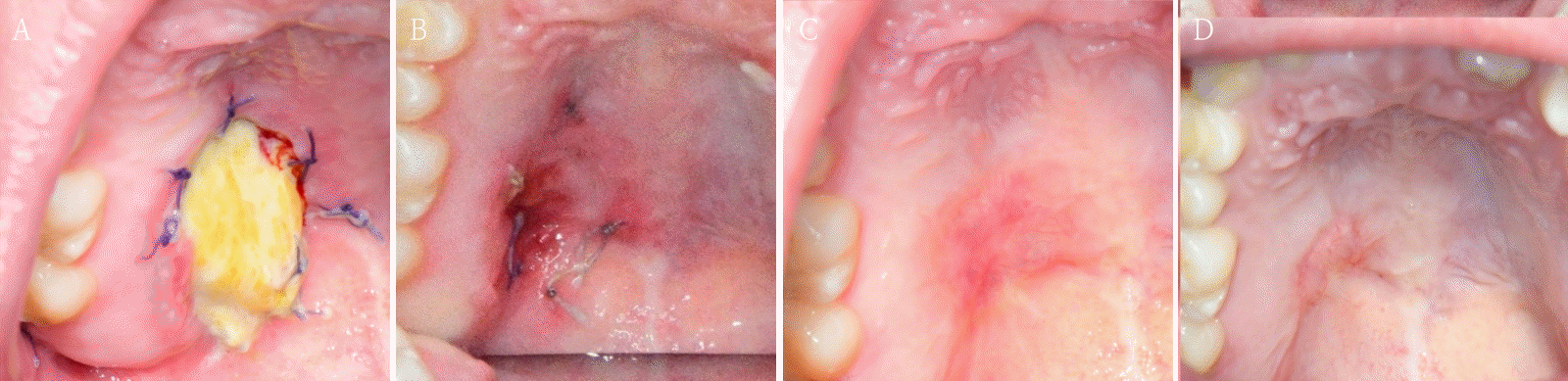
A 26-year-old female patient was referred to our clinic for definitive diagnosis and treatment due to bulging lesion on the right posterior palatal region. The patient reported being aware of the lesion for approximately one month. Notable swelling was observed in the right posterior hard palate. Upon palpation, a firm swelling exhibiting slight elasticity was noted, without any evidence of fluctuation. The lesion did not extend beyond the palatal midline and measured approximately 2.5 cm x 2 cm x 2 cm. There was no color change on the overlying mucosa and the surface of the lesion was smooth without ulcers (Fig. 1A). It revealed no pain, and aspiration yielded no content. Adjacent teeth showed no clinical features such as pain or discomfort, and the palate involving the lesion exhibited no sensory abnormalities.A facial CT was performed, which revealed a 16 mm x 12 mm x 12 mm radiolucent cystic lesion with central faint high-density content (Figs. 1B and C). Incision biopsy was also done. Histopathological results confirmed the diagnosis of pleomorphic adenoma. Considering the possibility of recurrence, it was decided to exercise both the lesion and the overlying mucosa under general anesthesia. For the surgical defect that may arise postoperatively, a buccal fat pad free graft was designated as the reconstructive method. Under general anesthesia, a mass excision in the right palate with a buccal fat pad free graft was performed. The lesion with overlying mucosa was excised using an electrocautery device. The size of the excised lesion was approximately 2.5 cm x 2 cm x 2 cm. The border of the lesion was clear without adhesion. bony erosion of the palatal region was observed at the lesion site, with no evidence of maxillary sinus perforation. The posterior area of the right maxillary second molar was incised, and the dissection was performed to expose the buccal fat pad. A portion of the exposed fat pad was harvested and grafted into an excised defect as a free graft. The grafted buccal fat pad was sutured to the surrounding mucosa (Fig. 2). A prefabricated palatal shell was placed to protect the surgical field. Histopathological result of the excised lesion confirmed pleomorphic adenoma, with tumor-free resection margins. During the hospitalization period, the grafted buccal fat pad remained stable without evidence of complications.Three weeks postoperatively, the grafted buccal fat pad was observed to have remained viable without necrosis or infection. One month postoperatively, epithelialization of the grafted fat pad was noted, and the sutures were removed. At the two-month follow-up, the epithelialization of the grafted site had progressed without any complications. Finally at the eight-month follow-up, there was no recurrence. More follow up will be needed (Fig. 3).
- Discussion
- Discussion
Pleomorphic adenoma is a benign mixed tumor made up of myoepithelial and epithelial cells, which are encased in a fibrous capsule that isolates them from adjacent tissues. It represents the most prevalent benign neoplasm of the salivary glands, accounting for approximately 40–50 % of all salivary gland tumors. It most frequently arises in the parotid gland. Among minor salivary glands, the palate is the most involved site, with pleomorphic adenoma representing 40–70 % of cases. The tumor typically occurs in adults between the third and fifth decades of life and demonstrates a slight female predilection, although it can present across a broad age range [2]. Pleomorphic adenoma generally tends to grow slowly without pain, and surgical excision is commonly considered as the treatment of choice. The recurrence rate of the lesion after surgery varies from 0.5 % to 10 %, and the causes of recurrence may include inadequate diagnosis before surgery and improper surgical methods [3]. Pleomorphic adenoma located in the palatal region is typically surgically excised, and as in this case, buccal fat pad free grafts can be utilized to address the surgical defect. Autologous buccal fat pad graft was first introduced by Peter Egyedi in 1977 [4], successfully closing oroantral and oronasal fistulas through pedicled buccal fat pad graft. The buccal fat pad is anatomically located within masticatory space and is abundantly supplied with blood from three arteries: the temporal artery, the buccal artery, and the superior posterior alveolar artery [5]. The volume of the buccal fat pad averages 10.2 ml in males and 8.9 ml in females. The average thickness is approximately 6 mm, and the average weight is 9.7 g. Autologous buccal fat pad graft can be divided into two techniques: pedicled buccal fat pad graft and buccal fat free graft. Pedicled buccal fat pad graft is primarily utilized for oroantral fistula closure, reconstruction of defects subsequent to surgery for medication-related osteonecrosis of the jaw (MRONJ) in the posterior region, and reconstruction of defects resulting from the excision of palatal tumors. Several advantages of this technique, including high success rate, rich blood supply, rapid epithelialization, and a low incidence of complications have been demonstrated though various studies and clinical cases. However, this technique is not applicable when the defect is located in the anterior maxilla or when the volume of the defect is large, as it may lead to vestibule obliteration or trismus due to tissue contraction [6]. In order to overcome these limitations, buccal fat free graft has been introduced. It allows for easier manipulation and transfer of adipose tissue, enabling the use of larger volumes of fat tissue for reconstruction of more extensive defects which could not be addressed by the pedicled buccal fat pad graft. It also enables acquisition of a larger volume of fat tissue and facilitates manipulation without the need to pass through the teeth. Therefore, buccal fat free graft was considered an appropriate approach for this case and yielded favorable results. Moreover, buccal fat free graft can be utilized in various patients with conditions such as MRONJ, oral cancer, and cleft lip and palate. according to a case report by de Castro et al. in 2015 [7], buccal fat free graft was successfully performed to reconstruct a defect in the anterior maxilla following cleft lip and palate surgery, leading to successful closure of an oronasal fistula. According to a case report by Cheng et al. in 2024 [8], buccal fat free graft was performed on eight patients who underwent saucerization surgery due to MRONJ. All patients had extensive defects with poor bone vascularity; however, they all exhibited successful healing outcomes following buccal fat free graft. Similarly, a study by Kablan and Laster in 2016 [9], reported the successful reconstruction of a defect created after the removal of a pleomorphic adenoma on the palatal side using free buccal fat pad grafting. In his study, epithelialization of the grafted adipose tissue was observed 4 to 6 weeks post-surgery, with complete histological healing achieved within 3 to 4 months. Several complications associated with using the buccal fat pad for defect repair have been reported in the literature, including necrosis, failure, hematoma, infection, facial nerve injury, arterial bleeding, donor site morbidity, trismus, wound dehiscence, and excessive granulomatous tissue formation [3,7]. This report presented a case of a pleomorphic adenoma located in the posterior maxillary region of the right posterior palate, which exhibited bony invasion. To utilize the buccal fat pad for the surgical defect, it must transverse the molar area, which can lead to dysfunctions such as trismus and chewing, necrosis; therefore, a free graft was performed instead of a pedicled flap. In this case, the buccal fat was transplanted without complications, leading to successful epithelialization and maintained without recurrence. With careful case selection in the future, it is anticipated that buccal fat pad free grafts can be successfully incorporated for intraoral defects. This report highlighted the successful application of a buccal fat pad free graft in the reconstruction of a large intraoral defect. The donor site exhibited uneventful healing, and the grafted fat tissue demonstrated rapid epithelialization, ultimately resulting in functionally and aesthetically favorable outcomes. Importantly, the buccal fat pad achieved successful engraftment and tissue regeneration in the absence of a vascular pedicle, thereby supporting the potential utility of free fat grafting as a viable technique for intraoral defect reconstruction.
- NOTES
- NOTES
Fig. 1.
A. A round, protruding lesion is observed in the posterior region of the right hard palate, with no associated symptoms such as pain. B and C. Facial CT images show no significant changes in size or bone destruction compared to the previous examination.
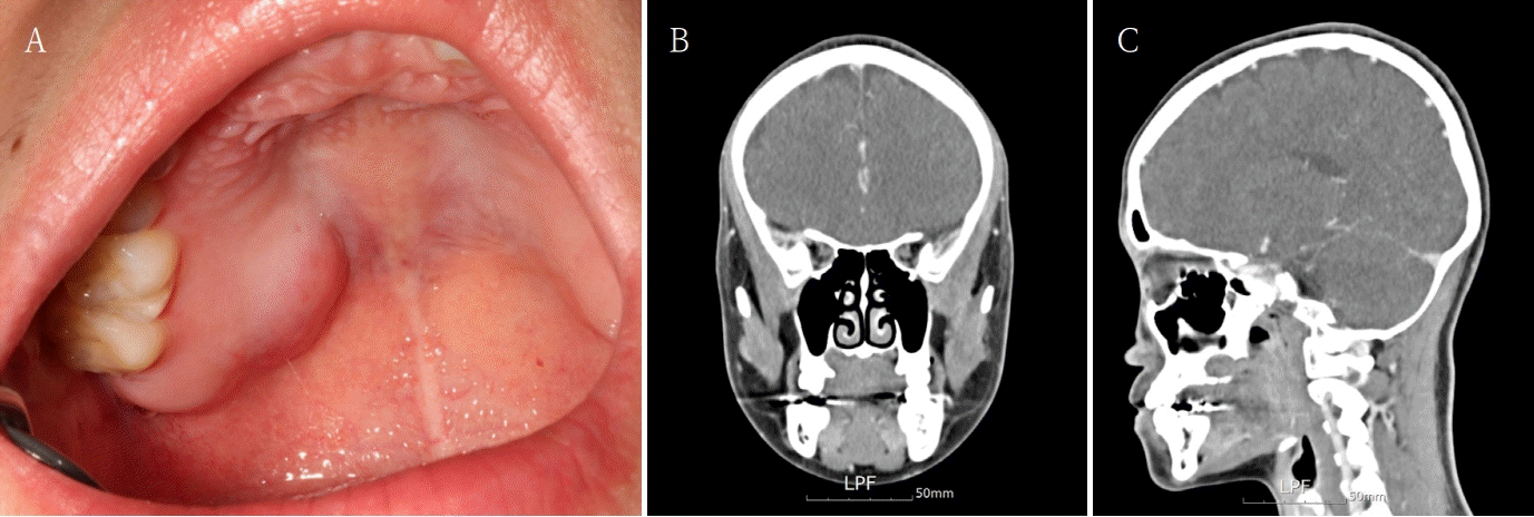
Fig. 2.
A. A well-defined lesion in the posterior region of the right hard palate before surgical intervention. B. The excised specimen obtained during surgical resection. C. Surgical site following complete excision of the lesion, exposing the underlying defect (bony erosion but no sinus perforation). Harvesting of the buccal fat pad from the buccal region for defect reconstruction. D. The buccal fat pad free graft placed within the defect site to promote wound healing and tissue regeneration.
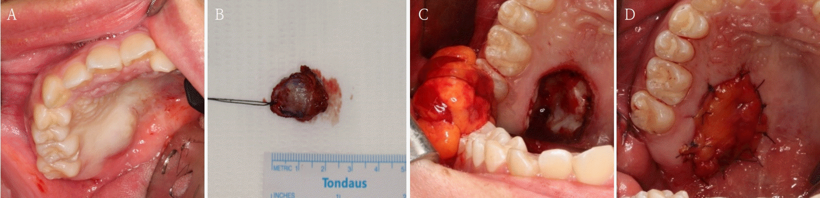
Fig. 3.
Post operation state. A. Post-operative day 20, No infection and necrosis. B. Post-operative day 1 month, epithelization on op site. C. Post-operative day 2 months, good healing state. D. Post-operative day 8 months, No recurrence.
- REFERENCES
- REFERENCES
- 1. Jain M.K., Ramesh C., Sankar K., Lokesh Babu K.T.. Pedicled buccal fat pad in the management of oroantral fistula: a clinical study of 15 cases. Int J Oral Maxillofac Surg 2012; 41: 1025–1029.
[Article] [PubMed]2. Yousra Z, Saliha C. Pleomorphic adenoma of hard palate: a case report. Pan Afr Med J 2021; 38: 146.
[Article] [PubMed] [PMC]3. Sheereen S, Manva MZ, Sheereen S, Patil NN, Abdelrahim RK, Afroz MM. Pleomorphic adenoma in salivary glands: insights from a 100-patient analysis. J Oral Maxillofac Pathol 2024; 28: 42–8.
[Article] [PubMed] [PMC]4. Egyedi P. Utilization of the buccal fat pad for closure of oro-antral and/or oro-nasal communications. J Maxillofac Surg 1977; 5: 241–4.
[Article] [PubMed]5. Favero G, van Noorden CJ, Rezzani R. The buccal fat pad: a unique human anatomical structure and rich and easily accessible source of mesenchymal stem cells for tissue repair. Bioengineering (Basel) 2024; 11: 968.
[Article] [PubMed] [PMC]6. Kumar D, Rattan V, Rai S, Yadav S, Sahu GR. Reconstruction of anterior maxillary defect with buccal pad fat after excision of melanotic neuroectodermal tumor of infancy. Ann Maxillofac Surg 2015; 5: 234–6.
[Article] [PubMed] [PMC]7. de Castro CH, de Souza LN, Fernandes Santos Melo M. Use of the buccal fat pad as free graft for closure of oronasal fistula in a cleft palate patient. J Craniofac Surg 2015; 26: e14–6.
[Article] [PubMed]8. Cheng JW, Hsiao KY, Chen MY, Huang TT, Chen KC. Free buccal fat pad graft for the bone defect filling of medication-related osteonecrosis of the jaws: a novel surgical approach. J Dent Sci 2024; 19: 1846–9.
[Article] [PubMed] [PMC]9. Kablan F, Laster Z. The use of free fat tissue transfers from the buccal fat pad to obtain and maintain primary closure and to improve soft tissue thickness at bone-augmented sites: technique presentation and report of case series. Int J Oral Maxillofac Implants 2014; 29: e220–31.
[Article] [PubMed]