


Ischemic necrosis of the tongue in a patient following vasopressor agents use and prolonged endotracheal intubation: A case report
-
Seung-Heon Bae+
 , Sung-Tak Lee+, Jin-wook Kim*
- Received March 27, 2025 Revised June 23, 2025 Accepted July 28, 2025
- ABSTRACT
-
Ischemic necrosis of the tongue is rare condition due to its rich vascular supply. No cases have been reported in Korea to date. A 55-year-old male with a history of dyslipidemia and fatty liver presented with abdominal pain and vomiting. Despite fluid resuscitation, he developed severe hypotension, requiring vasopressor support and mechanical ventilation. During his intensive care unit stay, the patient exhibited signs of disseminated intravascular coagulation, with progressive skin necrosis and thrombocytopenia. Upon extubation, oral examination revealed widespread mucosal discoloration and blood clots, and ischemic necrosis on dorsal surface of the tongue was confirmed. This case highlights the potential risk of tongue ischemia as a rare but serious complication of multifactorial cause including prolonged vasopressor use and endotracheal intubation. Further studies are needed to better understand its pathophysiology and improve management strategies.
- Introduction
- Introduction
Vasopressors play a crucial role in the management of refractory shock, which is defined as hypotension that remains unresponsive to intravenous fluid resuscitation alone [1]. However, their prolonged use carries the risk of end-organ hypoperfusion, which can result in peripheral ischemia including upper and lower extremities affecting future functional status and quality of life [2,3]. Although the tongue is considered a peripheral organ, ischemic necrosis of the tongue is rarely reported. This rarity is attributed to its rich vascular supply, primarily from the lingual and submandibular arteries, which typically ensures adequate perfusion even in compromised systemic conditions [4]. Most cases of ischemic necrosis of the tongue are associated with giant cell arteritis [5]. However, although rarely reported, other systemic conditions such as sepsis and cardiogenic shock have also been implicated in this complication [6,7]. To the authors’ knowledge, no cases of ischemic necrosis of the tongue have been reported in South Korea to date. This study aims to report a case of ischemic necrosis of the tongue in a patient following usage of vasopressor agents and prolonged endotracheal intubation.
- Case Report
- Case Report
A 55-year-old man with a history of dyslipidemia and fatty liver presented to Kyungpook National University Hospital emergency center on May 10, 2023, with complaints of abdominal pain and vomiting that began the day prior. The patient reported significant alcohol consumption, including two bottles of alcohol daily for the past month and at least one bottle per meal over the previous 10 days. Initial laboratory evaluations revealed stage 3 acute kidney injury and severe metabolic acidosis, prompting the initiation of continuous renal replacement therapy (CRRT). Upon arrival, he was alert but exhibited hypotension (105/48 mmHg) and hypoxemia (SpO₂ 80%). Fluid resuscitation (125 cc/hr) and supplemental oxygen (15 L/min) were administered. However, within two hours, his condition rapidly deteriorated, with a decline in mental status to a semi-comatose state, blood pressure dropping to 59/27 mmHg, and oxygen saturation further decreasing to 71%. The patient was subsequently admitted to the intensive care unit (ICU), requiring continuous vasopressor support and mechanical ventilation via endotracheal intubation. Norepinephrine (Norpin 0.1% 10ml) mixed with dextrose infusion was initiated at 10 cc/hr but showed no significant improvement, necessitating an increase to 20 cc/hr. Vasopressin 20u/1ml was mixed with Normal saline and infusion was initiated at 5cc/hr. Epinephrine mixed with Normal saline was concurrently administered at 10 cc/hr. Vasopressor support continued for six days post-admission. On the second day of admission, tenderness and skin discoloration developed in the limbS region, followed by bullae formation and extensive skin detachment by May 12 (Fig. 1). Laboratory findings indicated disseminated intravascular coagulation (DIC), with markedly elevated d-dimer levels (>20), severe thrombocytopenia (platelet count 15 × 10⁹/L), hypofibrinogenemia (78 mg/dL), prolonged prothrombin time (23 seconds), activated partial thromboplastin time (53.8 seconds), and reduced antithrombin III (35.1%). Abdominal computed tomography (CT) revealed a hypoattenuated liver lesion, suggesting multiple organ failure. Antithrombin III injections were administered as part of DIC management. On the 9th day of admission, as the patient’s vital signs improved, he was transferred from the ICU to a general ward. However, progressive skin detachment continued, and dry gangrene of the right second finger was noted (Fig. 2). Vascular surgery consultation confirmed inotrope-induced necrosis. On the following day, endotracheal intubation was removed. Oral examination post-extubation revealed widespread mucosal discoloration and blood clots affecting the palate, gingiva, and tongue (Fig. 3). On the 14th day of admission, the department of oral and maxillofacial surgery was consulted for oral care. With no active bleeding, conservative management using chlorhexidine dressings was initiated. While mucosal discoloration of the palate and gingiva improved, tongue discoloration worsened progressively. On June 4, necrosis of the tongue was confirmed, leading to re-evaluation by the oral and maxillofacial surgery team. Passive debridement and hydrogen peroxide dressings were performed (Fig. 4). The patient was discharged on June 20 and returned for outpatient follow-up two days later. Residual necrotic tissue was removed, and at the two-week postoperative follow-up, healing was favorable with no recurrence. At a follow-up evaluation 1 year and 4 months later, no additional complications were observed (Fig. 5).
- Discussion
- Discussion
The tongue is a well-vascularized end organ, primarily receiving its rich blood supply from the lingual artery, a branch of the external carotid artery, with additional contributions from the facial and pharyngeal arteries [7,8]. Due to this extensive vascular network, ischemic necrosis of the tongue is extremely rare. The most commonly reported cause is giant cell arteritis (GCA), which typically presents unilaterally, with bilateral cases being exceptionally uncommon [5]. Laboratory findings in cases of GCA include elevated erythrocyte sedimentation rate (ESR), plasma fibrinogen, and alpha-2 globulin levels [9]. However, in this case, plasma fibrinogen was decreased and alpha-2 globulin remained within the normal range, and the patient exhibited ischemia on central surface of the tongue, making GCA an unlikely cause.In this case, the patient experienced prolonged use of vasopressor agents due to severe hypotension secondary to shock. Norepinephrine is a catecholamine and a potent α1-adrenergic receptor agonist that increases vascular resistance by inducing vasoconstriction. It also acts as a β1-adrenergic receptor agonist, enhancing myocardial muscle fiber contractility and increasing heart rate. Norepinephrine is considered the first-line agent for hypotension in shock [10]. However, prolonged use has been associated with ischemic necrosis of peripheral tissues [11]. While the exact dosage and conditions leading to tongue necrosis remain unclear, vasopressor agents are recognized contributors to ischemic complications [3,12]. For this patient, vasopressin was additionally used. Vasopressin, first reported in 1926 [13], is an antidiuretic hormone that promotes water reabsorption in the kidneys and induces vasoconstriction, thereby increasing blood pressure. A meta-analysis report conducted in 2019 suggested that the use of vasopressin, either alone or in combination, is associated with a higher risk of digital ischemia compared to norepinephrine [14]. This patient had additional risk factors, including acute kidney injury (AKI) and DIC, both of which are known to increase susceptibility to peripheral ischemia, even at low-dose infusion [15]. Notably, this patient also developed inotrope-induced necrosis of the second finger, supporting the hypothesis that vasopressor therapy played a significant role.The ischemic necrosis of the tongue is thought to result from vascular compromise, with a combination of multiple contributing factors rather than a single cause [7]. While vasopressor agent usage was a primary factor, other considerations must be addressed. As the patient has undergone prolonged endotracheal intubation, persistent mechanical pressure on the tongue may have caused continuous edema. This factor contribute to compromising of blood flow resulting in exacerbation of peripheral ischemia [8]. In this case, laboratory tests confirmed that the patient met the International Society on Thrombosis and Hemostasis (ISTH) diagnostic criteria (Table 1) for DIC, a condition marked by systemic hypercoagulability leading to both microvascular and macrovascular thrombosis, ultimately resulting in multiple organ dysfunction syndrome [16]. Multiple organ dysfunction typically affects end-organs first, including the limbs, as infarcted distal regions deprived of blood supply fail to undergo inflammatory changes, leading to tissue ischemia [17]. Despite being an end-organ, the tongue’s rich vascular supply usually prevents necrosis under normal conditions. Nevertheless, cases of ischemic necrosis of the tongue associated with DIC have been documented [18]. Despite its rich vascularization, the tongue can still be affected under extreme ischemic conditions.Several reports suggest that cardiogenic shock and septic shock can lead to tongue necrosis, although the exact mechanism remains unclear [6,7]. One study hypothesized that severe shock leads to circulatory volume depletion, prioritizing blood flow to the internal carotid artery at the expense of the external carotid system, which could compromise blood supply to the tongue [6].Conservative treatment is often the preferred approach for ischemic tongue necrosis. Necrotic tissue undergoes autoamputation over time, with healing occurring by secondary intention, which may result in functional deficits affecting speech and swallowing [4]. In this case, conservative management was successful, with only minor necrotic tissue removal performed post-discharge. The patient was offered rehabilitation therapy to address residual speech difficulties.This case suggests that ischemic necrosis of the tongue is a rare condition but can occur as a complication associated with prolonged use of vasopressor agents. Multiple contributing factors may interact in its development, including mechanical compression from prolonged intubation, DIC, and circulatory volume depletion after shock. Given the rarity of this condition, clinicians should remain vigilant in recognizing potential risk factors and implementing timely interventions. Further research is necessary to examine the underlying mechanisms of ischemic necrosis of the tongue and optimize treatment approaches for affected patients.
- NOTES
- NOTES
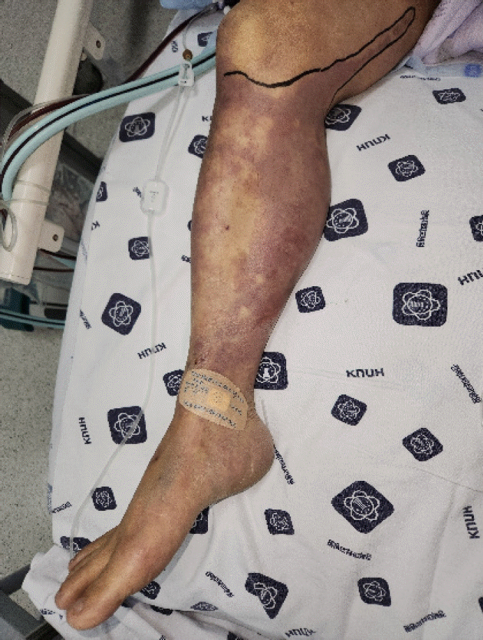
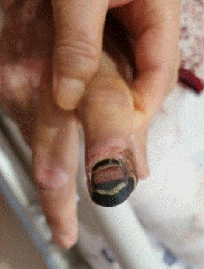
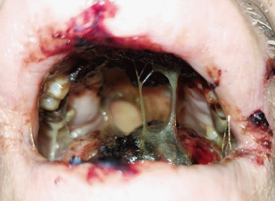
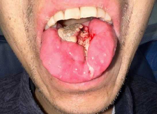
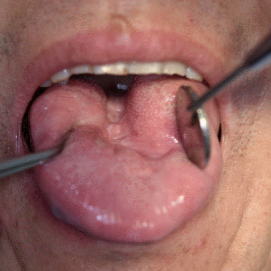
Table 1.
Diagnostic criteria for disseminated intravascular coagulation (DIC) by International Society on Thrombosis and Hemostasis [19]
- REFERENCES
- REFERENCES
- 1. Attallah N, Hassan E, Jama AB, Jain S, Ellabban M, Gleitz R, et al. Management of vasopressor-induced acute limb ischemia (VIALI) in septic shock. Cureus 2022; 14: e33118.
[Article] [PubMed] [PMC]2. Martin C, Viviand X, Leone M, Thirion X. Effect of norepinephrine on the outcome of septic shock. Crit Care Med 2000; 28: 2758–65.
[Article] [PubMed]3. Livesey M, Jauregui JJ, Hamaker MC, Pensy RA, Langhammer CG, Eglseder WA. Management of vasopressor induced ischemia. J Orthop 2020; 22: 497–502.
[Article] [PubMed] [PMC]4. McGoldrick DM, Khan I, Cotter CJ. Ischaemic necrosis of the tongue. BMJ Case Rep 2015; 2015: bcr2014208330.
[Article] [PubMed] [PMC]5. Brodmann M, Dorr A, Hafner F, Gary T, Pilger E. Tongue necrosis as first symptom of giant cell arteritis (GCA). Clin Rheumatol 2009; 28 Suppl 1: S47–9.
[Article] [PubMed]6. Roman BR, Immerman SB, Morris LG. Ischemic necrosis of the tongue in patients with cardiogenic shock. Laryngoscope 2010; 120: 1345–9.
[Article] [PubMed] [PMC]7. Cho J, Sung K, Lee D. Ischemic necrosis of the tongue in surgical patients with septic shock: a case report. BMC Surg 2016; 16: 48.
[Article] [PubMed] [PMC]8. Adegbite NA, Avery C, Rajaram K, Mohamed Ahmed M. Tongue necrosis: a rare complication of prolonged oral intubation. J Surg Case Rep 2019; 2019: rjz284.
[PubMed] [PMC]9. Llorente Pendás S, De Vicente Rodríguez JC, González García M, Junquera Gutierrez LM, López Arranz JS. Tongue necrosis as a complication of temporal arteritis. Oral Surg Oral Med Oral Pathol 1994; 78: 448–51.
[Article] [PubMed]10. Dellinger RP, Levy MM, Rhodes A, Annane D, Gerlach H, Opal SM, et al. Surviving sepsis campaign: international guidelines for management of severe sepsis and septic shock: 2012. Crit Care Med 2013; 41: 580–637.
[PubMed]11. Ruslan MA, Baharuddin KA, Noor NM, Yazid MB, Noh AY, Rahman A. Norepinephrine in septic shock: a systematic review and meta-analysis. West J Emerg Med 2021; 22: 196–203.
[PubMed] [PMC]12. Noordally SO, Sohawon S, Duttmann R, Gottignies P, Devriendt J. Tongue necrosis as a complication of vasoconstrictor agents in the intensive care setting. Intern Emerg Med 2011; 6: 183–5.
[Article] [PubMed]13. Geiling E, Campbell D. Variations in blood pressure induced by repeated injections of extracts of the posterior lobe of the pituitary gland. J Pharmacol Exp Ther 1926; 29: 449–60.
[Article]14. Nagendran M, Russell JA, Walley KR, Brett SJ, Perkins GD, Hajjar L, et al. Vasopressin in septic shock: an individual patient data meta-analysis of randomised controlled trials. Intensive Care Med 2019; 45: 844–55.
[Article] [PubMed]15. Cho AR, Kim JI, Kim EJ, Son SM. Skin necrosis after high dose vasopressor infusion in septic shock: two case reports. Acute Crit Care 2012; 27: 182–6.
[Article]16. Costello RA, Leslie SW, Nehring SM. Disseminated intravascular coagulation. In: StatPearls; Treasure Island (FL): 2025.17. Bick RL, Arun B, Frenkel EP. Disseminated intravascular coagulation. clinical and pathophysiological mechanisms and manifestations. Haemostasis 1999; 29: 111–34.
[PubMed]